. 2011 Jun 3;10(6):2785-96.
doi: 10.1021/pr200042u. Epub 2011 Apr 29.

Mechlorethamine-induced DNA-protein cross-linking in human fibrosarcoma (HT1080) cells

Affiliations

Mechlorethamine-induced DNA-protein cross-linking in human fibrosarcoma (HT1080) cells

Erin D Michaelson-Richie et al. J Proteome Res. .

Abstract

Antitumor nitrogen mustards, such as bis(2-chloroethyl)methylamine (mechlorethamine), are useful chemotherapeutic agents with a long history of clinical application. The antitumor effects of nitrogen mustards are attributed to their ability to induce DNA-DNA and DNA-protein cross-links (DPCs) that block DNA replication. In the present work, a mass spectrometry-based methodology was employed to characterize in vivo DNA-protein cross-linking following treatment of human fibrosarcoma (HT1080) cells with cytotoxic concentrations of mechlorethamine. A combination of mass spectrometry-based proteomics and immunological detection was used to identify 38 nuclear proteins that were covalently cross-linked to chromosomal DNA following treatment with mechlorethamine. Isotope dilution HPLC-ESI(+)-MS/MS analysis of total proteolytic digests revealed a concentration-dependent formation of N-[2-(S-cysteinyl)ethyl]-N-[2-(guan-7-yl)ethyl]methylamine (Cys-N7G-EMA) conjugates, indicating that mechlorethamine cross-links cysteine thiols within proteins to N-7 positions of guanine in DNA.


Figures
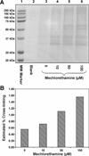
Figure 1
Figure 1
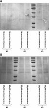
Concentration-dependent formation of DNA-protein cross-links in HT1080 cells treated with mechlorethamine. Cells were treated with 0–100 µM mechlorethamine for 3 h, and chromosomal DNA containing cross-linked proteins was isolated by modified phenol/chloroform extraction in the presence of proteasome inhibitors. Proteins (from 15 µg DNA) were released from DNA by thermal hydrolysis, separated by SDS-PAGE, and visualized by staining with SimplyBlue SafeStain (A). Densitometric analysis of protein bands in the 25–250 kDa molecular weight region in comparison with total protein extracts was used to approximate the extent of DNA-protein cross-linking (B).
Figure 2
Figure 2
SDS-PAGE analysis of samples employed in the proteomics studies of mechlorethamine-induced DPCs. HT1080 cells (~107) were treated with 0 (A) or 25 µM mechlorethamine for 3 h (B). Following modified phenol/chloroform extraction of DNA in the presence of proteasome inhibitors and thermal hydrolysis to release proteins, the cross-linked proteins were separated by 12% SDS-PAGE and visualized by staining with SimplyBlue SafeStain. Proteins present in the 20 – 250 kDa molecular weight range were excised from the gel, subjected to in-gel tryptic digestion, and analyzed by HPLC-ESI+-MS/MS.
Figure 3
Figure 3
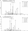
Representative HPLC-ESI+-MS/MS spectra of tryptic peptides used in the identification of DPCs involving matrin-3 (A), and zinc finger Ran-binding domain-containing protein 2 (B).
Figure 4
Figure 4
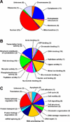
GO annotations for proteins involved in mechlorethamine-induced DPC formation in human HT1080 cells: cellular distributions (A), molecular functions (B), and biological processes (C). The number of proteins in each category is labeled on the charts.
Figure 5
Figure 5
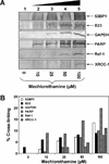
Western blot analysis of mechlorethamine-induced DPCs in HT1080 cells. Following treatment with 0 (lane 1), 10 (lane 2), 25 (lane 3), or 50 µM mechlorethamine (lane 4), DNA and covalently cross-linked proteins were isolated by phenol/chloroform extraction. Samples were normalized for DNA content, and proteins from 15 µg DNA were released by thermal hydrolysis, separated by SDS-PAGE, and transferred to nitrocellulose membranes. Western blotting was performed using primary antibodies specific for 53BP1, B23, GAPDH, PARP, Ref-1, and XRCC-1 (A). The efficiency of DPC formation in the presence of mechlorethamine was estimated by densitometric analysis of protein bands in DPC samples and a whole cell protein lysate control (B).
Figure 6
Figure 6
Western blot analysis of mechlorethamine-induced AGT-DNA cross-links in Chinese hamster ovary (CHO) cells expressing human AGT ptotein. Following treatment with 0 (lane 1), 1 (lane 2), 5 (lane 3), 10 (lane 4), 25 (lane 5) or 50 µM mechlorethamine (lane 6) for 3 hours, DNA was isolated by modified phenol/chloroform extraction in the presence of proteasome inhibitors. DNA aliquots (15 µg) were subjected to thermal hydrolysis to release proteins, which were separated by SDS-PAGE and transferred to nitrocellulose membranes. Western blotting was performed using a primary antibody against AGT.
Figure 7
Figure 7
HPLC-ESI+-MS/MS analysis of Cys-N7G-EMA conjugates in total proteolytic digests of mechlorethamine-induced DPCs. HT1080 cells were treated with mechlorethamine to induce DNA-protein cross-linking. Following extraction of DPC-containing chromosomal DNA, the cross-linked proteins were subjected to thermal and enzymatic hydrolysis to release amino acid-nucleobase conjugates. Digest mixtures were spiked with isotopically labeled internal standard (Cys-15N5-N7G-EMA) to enable the direct quantitation of Cys-N7G-EMA. Shown in the figure are extracted ion chromatograms corresponding to HT1080 cells incubated in the absence of mechlorethamine (negative control) (A); and samples treated with 10 µM mechlorethamine (B).
Figure 8
Figure 8
Concentration dependent formation of Cys-N7G-EMA in mechlorethamine-treated HT1080 cells. HT1080 cells were exposed to 0, 10, 25, 50, or 100 µM mechlorethamine for 3 h. Following extraction of DPC-containing chromosomal DNA, equal DNA amounts from each sample were subjected to thermal and enzymatic hydrolysis to release amino acid-nucleobase conjugates. The samples were subjected to offline HPLC to enrich for Cys-N7G-EMA prior to HPLC-ESI+-MS/MS analysis. Quantification of Cys-N7G-EMA was accomplished using isotope dilution with Cys-15N5-N7G-EMA. Error bars represent the standard error of three independent experiments.
Scheme 1
Scheme 1
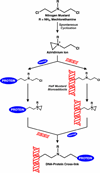
Formation of DPCs by antitumor nitrogen mustards
Scheme 2
Scheme 2
Strategy for the isolation and analysis of DPCs from mechlorethamine-treated mammalian cell cultures.

References

    1. Zhitkovich A, Voitkun V, Kluz T, Costa M. Utilization of DNA-protein cross-links as a biomarker of chromium exposure. Environ. Health Perspect. 1998;106 Suppl 4:969–974. - PMC - PubMed
    1. Barker S, Weinfeld M, Zheng J, Li L, Murray D. Identification of mammalian proteins cross-linked to DNA by ionizing radiation. J. Biol. Chem. 2005;280:33826–33838. - PubMed
    1. Murata-Kamiya N, Kamiya H. Methylglyoxal, an endogenous aldehyde, crosslinks DNA polymerase and the substrate DNA. Nucleic Acids Res. 2001;29:3433–3438. - PMC - PubMed
    1. Kloster M, Kostrhunova H, Zaludova R, Malina J, Kasparkova J, Brabec V, Farrell N. Trifunctional dinuclear platinum complexes as DNA-protein cross-linking agent. Biochemistry. 2004;43:7776–7786. - PubMed
    1. Ewig RA, Kohn KW. DNA-protein cross-linking and DNA interstrand cross-linking by haloethylnitrosoureas in L1210 cells. Cancer Res. 1978;38:3197–3203. - PubMed

Publication types

MeSH terms