. 2011 Apr 12:11:72.
doi: 10.1186/1471-2180-11-72.

Contribution of SecDF to Staphylococcus aureus resistance and expression of virulence factors

Affiliations

Contribution of SecDF to Staphylococcus aureus resistance and expression of virulence factors

Chantal Quiblier et al. BMC Microbiol. .

Abstract

Background: SecDF is an accessory factor of the conserved Sec protein translocation machinery and belongs to the resistance-nodulation-cell division (RND) family of multidrug exporters. SecDF has been shown in Escherichia coli and Bacillus subtilis to be involved in the export of proteins. RND proteins can mediate resistance against various substances and might be of relevance in antimicrobial therapy. The role of RND proteins in Staphylococcus aureus has not yet been determined.

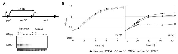
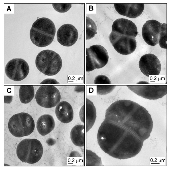
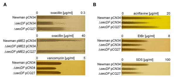
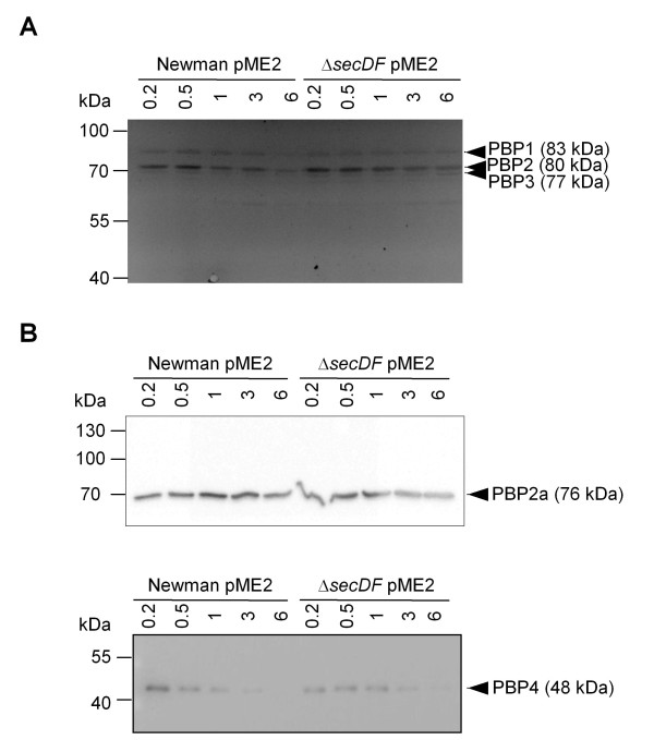
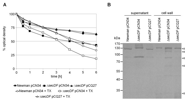
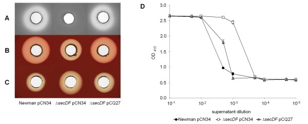
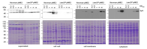
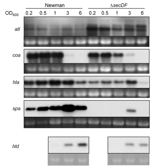
Results: Markerless deletion mutants were constructed to analyze the impact of the so far uncharacterized RND proteins in S. aureus. While the lack of Sa2056 and Sa2339 caused no phenotype regarding growth and resistance, the secDF mutant resulted in a pleiotropic phenotype. The secDF mutant was cold sensitive, but grew normally in rich medium at 37°C. Resistance to beta-lactams, glycopeptides and the RND substrates acriflavine, ethidium bromide and sodium dodecyl sulfate was reduced. The secDF mutant showed an aberrant cell separation and increased spontaneous and Triton X-100 induced autolysis, although the amounts of penicillin-binding proteins in the membrane were unchanged. The impact of secDF deletion on transcription and expression of specific virulence determinants varied: While coagulase transcription and activity were reduced, the opposite was observed for the autolysin Atl. A reduction of the transcription of the cell wall anchored protein A (spa) was also found. The accumulation of SpA in the membrane and lowered amounts in the cell wall pointed to an impaired translocation.

Conclusions: The combination of different effects of secDF deletion on transcription, regulation and translocation lead to impaired cell division, reduced resistance and altered expression of virulence determinants suggesting SecDF to be of major relevance in S. aureus. Thus SecDF could be a potential target for the control and eradication of S. aureus in the future.


Figures

Figure 1
Figure 1
Growth characteristics of the secDF mutant. (A) Genetic context of secDF in S. aureus and Northern blot analysis of secDF transcription during growth. Predicted promoter and terminators are depicted. Ethidium bromide-stained 16S rRNA is shown as an indication of RNA loading. (B) Growth of Newman and the secDF mutant carrying the empty vector pCN34, and of the complemented mutant secDF pCQ27 in LB broth at 37°C and 15°C, respectively.
Figure 2
Figure 2
Cell morphology. TEM pictures from thin sections of strains (A) Newman pCN34, (B and D) ΔsecDF pCN34 and (C) ΔsecDF pCQ27 during exponential phase (OD600 0.5).
Figure 3
Figure 3
Effect of secDF inactivation on resistance profiles. (A) Gradient plates with increasing concentrations of β-lactam and glycopeptide antibiotics. Oxacillin was tested with methicillin sensitive and methicillin resistant strain Newman, the latter carrying the plasmid pME2 containing the mecA gene. (B) Gradient plates with increasing concentrations of the RND substrates acriflavine, ethidium bromide and SDS.
Figure 4
Figure 4
PBP expression over growth. Strain Newman pME2, carrying mecA, and its secDF mutant were cultivated in LB and samples collected at the indicated OD600 were used to prepare membrane fractions. (A) Membranes were incubated with the fluorescent penicillin analogue Bocillin-FL. Bands corresponding to PBPs 1-3 are indicated. (B) Western blot analysis of membrane fractions using antibodies against PBP2a and PBP4, respectively.
Figure 5
Figure 5
Autolysis and zymogram. (A) Spontaneous and Triton X-100 (TX) induced autolysis was measured over time. (B) Autolysin zymography of protein extracts from supernatant and cell wall was performed using SDS-10% PAGE supplemented with S. aureus cell wall extract as a substrate. Dark bands show hydrolyzed cell wall and are indicated by triangles. Based on the work of Schlag et al. bands were assigned as follows in decreasing order: Pro-Atl (~130 kDa); Atl (~115 kDa); Atl-amidase (~84 kDa) or part of the propeptide (62-65 kDa); Sle1/Aaa (~33 kDa) [35].
Figure 6
Figure 6
Proteolysis and hemolysis of sessile and planktonic bacteria. Proteolytic and hemolytic activity was determined qualitatively by agar diffusion assay on skim milk, respectively sheep blood agar. Hemolytic activity was measured in diluted sheep blood. (A) Skim milk agar and (B) sheep blood agar with sessile bacteria. (C) Sheep blood agar with sterile-filtered supernatants of stationary phase planktonic bacteria. (D) Release of hemoglobin by stationary phase supernatants of planktonic bacteria. Representative data of three independent experiments are shown with standard deviations of technical triplicates.
Figure 7
Figure 7
Subcellular localization of SpA. Expression and localization of SpA was monitored in the Newman pME2 background during growth. Upper panels show Western blots of SpA. Longer exposure times were required for detection of SpA in cell membrane and cytoplasm. Bottom panels show Coomassie stained gels. Bands of stronger expression in the mutant are indicated by triangles.
Figure 8
Figure 8
Transcription of virulence factors. atl, coa, hla, spa and hld transcription was monitored over growth in strains Newman and ΔsecDF. Ethidium bromide-stained 16S rRNA is shown as an indication of RNA loading.

Similar articles

Cited by

References

    1. Sibbald MJJB, Ziebandt AK, Engelmann S, Hecker M, de Jong A, Harmsen HJM, Raangs GC, Stokroos I, Arends JP, Dubois JYF. et al.Mapping the pathways to staphylococcal pathogenesis by comparative secretomics. Microbiol Mol Biol Rev. 2006;70(3):755–788. doi: 10.1128/MMBR.00008-06. - DOI - PMC - PubMed
    1. Driessen AJM, Nouwen N. Protein translocation across the bacterial cytoplasmic membrane. Annu Rev Biochem. 2008;77(1):643–667. doi: 10.1146/annurev.biochem.77.061606.160747. - DOI - PubMed
    1. Pogliano JA, Beckwith J. SecD and SecF facilitate protein export in Escherichia coli. EMBO J. 1994;13:554–561. - PMC - PubMed
    1. Duong F, Wickner W. The SecDFyajC domain of preprotein translocase controls preprotein movement by regulating SecA membrane cycling. EMBO J. 1997;16(16):4871–4879. doi: 10.1093/emboj/16.16.4871. - DOI - PMC - PubMed
    1. Nouwen N, Piwowarek M, Berrelkamp G, Driessen AJM. The large first periplasmic loop of SecD and SecF plays an important role in SecDF functioning. J Bacteriol. 2005;187(16):5857–5860. doi: 10.1128/JB.187.16.5857-5860.2005. - DOI - PMC - PubMed

Publication types

MeSH terms

LinkOut - more resources