. 2011 Apr 12:8:31.
doi: 10.1186/1742-2094-8-31.

Selective adenosine A2A receptor agonists and antagonists protect against spinal cord injury through peripheral and central effects

Affiliations

Selective adenosine A2A receptor agonists and antagonists protect against spinal cord injury through peripheral and central effects

Irene Paterniti et al. J Neuroinflammation. .

Abstract

Background: Permanent functional deficits following spinal cord injury (SCI) arise both from mechanical injury and from secondary tissue reactions involving inflammation. Enhanced release of adenosine and glutamate soon after SCI represents a component in the sequelae that may be responsible for resulting functional deficits. The role of adenosine A2A receptor in central ischemia/trauma is still to be elucidated. In our previous studies we have demonstrated that the adenosine A2A receptor-selective agonist CGS21680, systemically administered after SCI, protects from tissue damage, locomotor dysfunction and different inflammatory readouts. In this work we studied the effect of the adenosine A2A receptor antagonist SCH58261, systemically administered after SCI, on the same parameters. We investigated the hypothesis that the main action mechanism of agonists and antagonists is at peripheral or central sites.

Methods: Spinal trauma was induced by extradural compression of SC exposed via a four-level T5-T8 laminectomy in mouse. Three drug-dosing protocols were utilized: a short-term systemic administration by intraperitoneal injection, a chronic administration via osmotic minipump, and direct injection into the spinal cord.

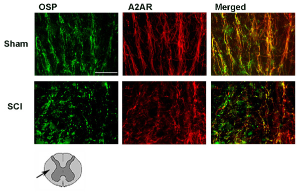
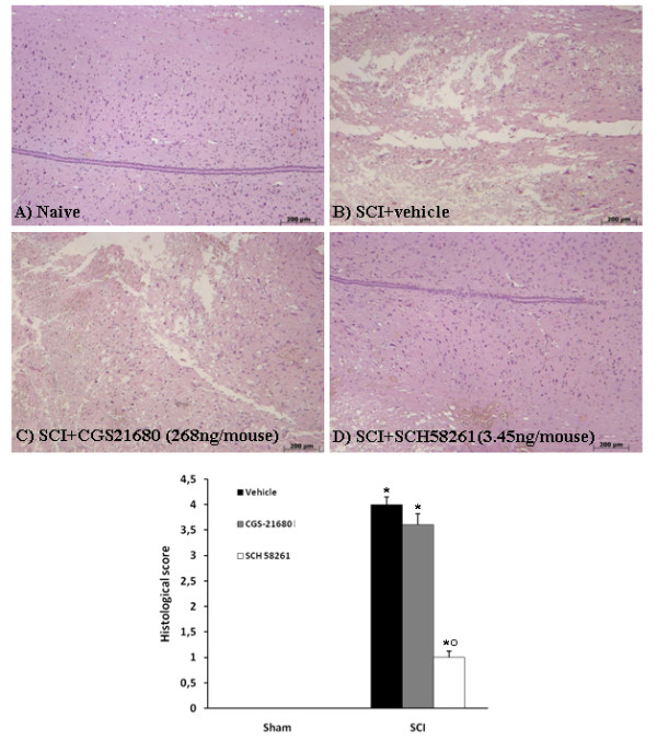
Results: SCH58261, systemically administered (0.01 mg/kg intraperitoneal. 1, 6 and 10 hours after SCI), reduced demyelination and levels of TNF-α, Fas-L, PAR, Bax expression and activation of JNK mitogen-activated protein kinase (MAPK) 24 hours after SCI. Chronic SCH58261 administration, by mini-osmotic pump delivery for 10 days, improved the neurological deficit up to 10 days after SCI. Adenosine A2A receptors are physiologically expressed in the spinal cord by astrocytes, microglia and oligodendrocytes. Soon after SCI (24 hours), these receptors showed enhanced expression in neurons. Both the A2A agonist and antagonist, administered intraperitoneally, reduced expression of the A2A receptor, ruling out the possibility that the neuroprotective effects of the A2A agonist are due to A2A receptor desensitization. When the A2A antagonist and agonist were centrally injected into injured SC, only SCH58261 appeared neuroprotective, while CGS21680 was ineffective.

Conclusions: Our results indicate that the A2A antagonist protects against SCI by acting on centrally located A2A receptors. It is likely that blockade of A2A receptors reduces excitotoxicity. In contrast, neuroprotection afforded by the A2A agonist may be primarily due to peripheral effects.


Figures

Figure 1
Figure 1
Effects of systemic SCH58261 treatment on histological alterations and on hind limb motor disturbance after SCI. Twenty-four hours after trauma, significant damage to the SC of untreated SCI-operated mice in the perilesional area was assessed by the presence of alterations of white matter (b). It is noteworthy that significant protection from the SCI was observed in tissue collected from SCH58261-treated SCI-injured mice (c). No significant alterations were observed in sections obtained in sham groups (a). Myelin structure was observed by Luxol fast blue staining as well by Weigert's and Oil red O staining. Twenty-four hours after injury in SCI-operated mice (e, h respectively) a significant loss of myelin was observed. In contrast, myelin degradation was attenuated (f, i respectively) in SCH58261-treated mice. No significant alterations were observed in sections obtained in sham groups (d, g respectively). This figure is representative of at least 3 experiments performed on different experimental days. The histological score (l) was evaluated by an independent observer. ND: not detectable. One-way ANOVA: *°P < 0.01 vs sham group and SCI+vehicle, respectively. The degree of motor disturbance was assessed every day until 10 days after SCI by BMS motor score (m). Systemic administration of SCH58261 reduced the motor disturbance starting from the fourth day after SCI (m). Values are shown as mean ± S.E., with 10 mice in each group. One-way ANOVA: *P < 0.01 vs SCI+vehicle.
Figure 2
Figure 2
Effects of systemic SCH58261-administration on inflammation parameters. A substantial increase in TNF-α (2b), Fas-L (2e) and PAR (2h) expression was found in SC tissue collected from SCI+vehicle-treated mice 24 hours after SCI, in comparison with sham-operated mice (2a, d, g respectively). TNF-α (2c), Fas-L (2f) and PAR (2i) levels were attenuated in the SCI+SCH58261 group in comparison to SCI+vehicle animals. Sections of SC from sham vehicle-treated mice did not stain for Bax (2l) whereas SC sections obtained from SCI mice exhibited positive staining (2m). SCH58261 reduced the degree of staining for Bax (2n). Spinal cord sections from sham vehicle-treated mice demonstrated Bcl-2 positive staining (2o), whereas in SCI control mice the staining was significantly reduced (2p). SCH58261 attenuated the loss of positive staining for Bcl-2 in the SC from SCI-subjected mice (2q).
Figure 3
Figure 3
Co-localization of adenosine A2A receptors with neurons (Nissl) in the gray matter of SC 24 h after injury. Double immunofluorescence was used to characterize the co-localization of A2A receptors (in red with Texas red) with neurons (in green with NeutroTracer green fluorescent Nissl stain) in sham-operated and SCI mice groups. The merged images show that A2A receptors are present on many neurons of the gray matter after SCI, but not all neurons expressed A2A receptors. The drawing under the figures shows localization of A2A receptors as indicated by boxes. Scale bar = 50 μm.
Figure 4
Figure 4
Co-localization of A2A receptors with astrocytes (GFAP) in white matter of SC 24 h after injury. Double immunofluorescence was used to characterize the co-localization of A2A receptors (in red with Texas red) with astrocytes (in green with fluorescein) in sham-operated and SCI mice groups. The merged images show that A2A receptors are present on astrocytes both before and after SCI. The co-localization is quite total. The drawing under figures shows that A2A receptors were identified in the white matter area as indicated by arrow. Scale bar = 10 μm.
Figure 5
Figure 5
Co-localization of A2A receptors with microglial cells (IBA1) in white matter of SC 24 h after injury. Double immunofluorescence was used to characterize the co-localization of A2A receptors (in red with Texas red) with microglia (in green with fluorescein) in sham-operated and SCI mice groups. The merged images show that A2A receptors are not present on activated microglial cells of the SC. The drawing under figures shows that A2A receptors were identified in the white matter area as indicated by arrow. Scale bar = 10 μm.
Figure 6
Figure 6
Co-localization of A2A receptors with oligodendrocyte myelinated bundles (OSP) in the white matter of SC 24 h after injury. Double immunofluorescence was used to characterize the co-localization of A2A receptors (in red with Texas red) with myelinated bundles (in green with fluorescein) in sham-operated and SCI mice groups. The merged images show that A2A receptors are present in myelinated bundles of the white matter after SCI. The drawing under figures shows that A2A receptors were identified in the white matter area as indicated by arrow. Scale bar = 50 μm.
Figure 7
Figure 7
Effects of systemic SCH58261- and CGS21680-administration on expression of A2A receptors. Western blot analysis shows that A2A receptors are detected in the SC of sham-operated animals and that they are substantially increased in SCI mice. SCH58261 and CGS21680 treatment significantly reduced SCI-induced A2A receptors expression. GAPDH was used as internal control. A representative blot of lysates obtained from each mice group is shown. The densitometric analysis is expressed as the mean ± SEM for samples from 10 mice from each group and is normalized by control protein (GAPDH) levels, and reported in bar graphs. One-way ANOVA: *P < 0.01 vs sham; # P < 0.05 vs SCI; # # P < 0.01 vs SCI.
Figure 8
Figure 8
Effect of systemic SCH58261-administration on JNK MAPK activation. Western blot analysis shows a significant increase in phospho-JNK MAPK (Thr183/Tyr185) 24 h after SCI. SCH58261, intraperitoneally administered three times within 24 h, reduced SCI-induced phospho-JNK MAPK levels. A representative blot of lysates obtained from each mice group is shown. The densitometric analysis is expressed as the mean ± SEM for samples from 10 mice from each group and is normalized by control protein (β-actin) levels, and reported in a bar graph. One-way ANOVA: *P < 0.01 vs sham; ##P < 0.05 vs SCI.
Figure 9
Figure 9
Effect of SCH58261 and CGS21680 treatment on histological alterations when centrally applied after SCI. Significant damage to the SC from SCI-operated mice in the perilesional area was assessed by the presence of edema as well as alteration of the white matter 24 h after injury (B). Notably, significant protection from SCI was observed in tissue collected from SCH58261-treated mice (D), whereas CGS21680, when locally applied on spinal cord tissue, did not protect against SCI (C). This figure is representative of at least 3 experiments performed on different experimental days. The histological score (d) was evaluated by an independent observer. ND: not detectable. *P < 0.01 vs sham; °P < 0.01 vs SCI.

Similar articles

Cited by

References

    1. Maegele M, Riess P, Sauerland S, Bouillon B, Hess S, McIntosh TK, Mautes A, Brockmann M, Koebke J, Knifka J, Neugebauer EA. Characterization of a new rat model of experimental combined neurotrauma. Shock. 2005;23:476–481. doi: 10.1097/01.shk.0000159929.87737.5c. - DOI - PubMed
    1. Profyris C, Cheema SS, Zang D, Azari MF, Boyle K, Petratos S. Degenerative and regenerative mechanisms governing spinal cord injury. Neurobiol Dis. 2004;15:415–436. doi: 10.1016/j.nbd.2003.11.015. - DOI - PubMed
    1. Hagg T, Oudega M. Degenerative and spontaneous regenerative processes after spinal cord injury. J Neurotrauma. 2006;23:264–280. - PubMed
    1. Linden J. Adenosine in tissue protection and tissue re generation. Mol Pharmacol. 2005;67:1385–1387. doi: 10.1124/mol.105.011783. - DOI - PubMed
    1. Norenberg MD, Smith J, Marcillo A. The pathology of human spinal cord injury: defining the problems. J Neurotrauma. 2004;21:429–440. doi: 10.1089/089771504323004575. - DOI - PubMed

Publication types

MeSH terms

Substances