. 2011 May 6;585(9):1281-6.
doi: 10.1016/j.febslet.2011.04.009. Epub 2011 Apr 8.

Recognition of an ERAD-L substrate analyzed by site-specific in vivo photocrosslinking

Affiliations

Recognition of an ERAD-L substrate analyzed by site-specific in vivo photocrosslinking

Ann Marie Stanley et al. FEBS Lett. .

Abstract

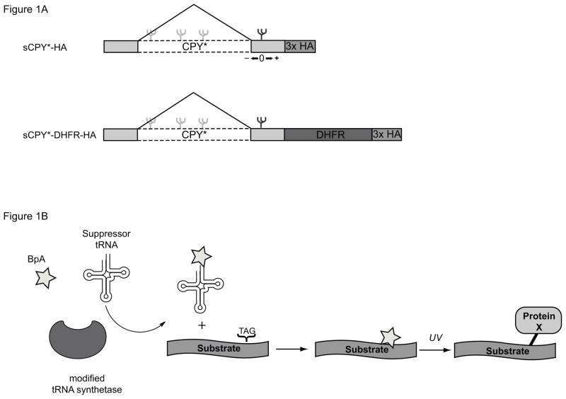
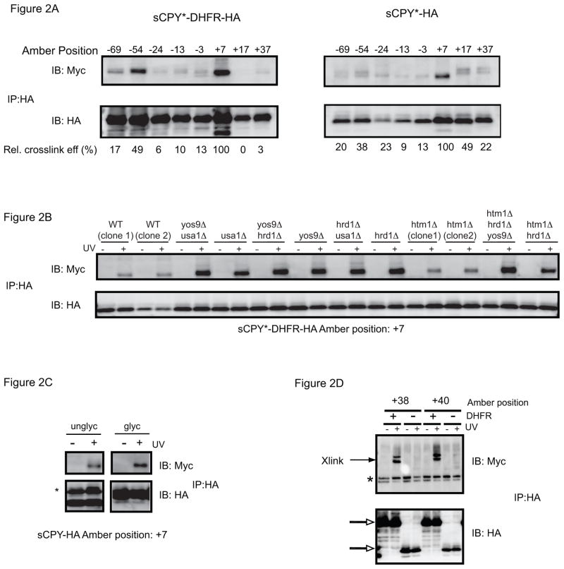
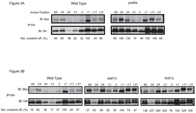
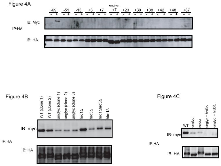
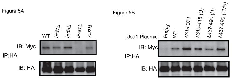
Misfolded, luminal endoplasmic reticulum (ER) proteins must be recognized before being degraded by a process called ERAD-L. Using site-specific photocrosslinking in Saccharomyces cerevisiae, we tested luminal interactions of a glycosylated ERAD-L substrate with potential recognition components. Major interactions were observed with Hrd3p. These are independent of the glycan and of other ERAD components, and can occur throughout the length of the unfolded substrate. The lectin Yos9p only interacts with a polypeptide segment distant from the degradation signal. Hrd3p may thus be the first substrate-recognizing component. Der1p appears to have a role in a pathway that is parallel to that involving Hrd3p.


Figures

Figure 1
Figure 1. Experimental design of in vivo site-specific photocrosslinking experiments
(A) Schematics of the ERAD-L substrates used. (B) Explanation of the in vivo photocrosslinking method. BpA, phenylalanine derivative containing a benzophenone.
Figure 2
Figure 2. Photocrosslinking to Hrd3p
(A) sCPY*-HA or sCPY*-DHFR-HA with amber codons at the indicated positions were expressed together with Hrd3p-13myc and subjected to in vivo photocrosslinking. Membranes were isolated and SDS-denatured proteins were immunoprecipitated with HA antibodies. The samples were analyzed by SDS-PAGE and immunoblotting with either HA or Myc antibodies. The numbers underneath the lanes give the relative crosslinking efficiency. For quantification, the intensity of the myc band was divided by that of the corresponding HA band, and normalized to the ratio determined for position +7. The comparison of crosslinking efficiency is only approximate, as the level of photocrosslinker incorporation may vary between positions. (B) sCPY*-HA with a photoreactive probe at position +7 was crosslinked to Hrd3p-13myc in cells lacking ERAD components. Where indicated, UV irradiation was omitted. (C) sCPY*-HA containing the N-glycosylation site (glyc) or lacking it (unglyc; mutation of N to A), both with a photoreactive probe at position +7, were crosslinked to Hrd3p-13myc. The asterisk indicates immunoglobulin light chain. (D) For comparison with (A), crosslinking to Hrd1p-13 myc was tested with sCPY*-HA or sCPY*-DHFR-HA containing a photoreactive probe at the indicated positions. The open arrows indicate the position of substrate, and the asterisk denotes non-specific bands reacting with Myc antibodies.
Figure 3
Figure 3. Photocrosslinking to Hrd3p in different ERAD mutants
(A) sCPY*-HA with amber codons at the indicated positions was expressed together with Hrd3p-13myc in wild type cells or cells lacking Yos9p. After in vivo photocrosslinking, membranes were isolated and SDS-denatured proteins were immunoprecipitated with HA antibodies. The samples were analyzed by SDS-PAGE and immunoblotting with either HA or Myc antibodies. The numbers underneath the lanes give the relative crosslinking efficiency. For quantification, the intensity of the myc band was divided by that of the corresponding HA band, and normalized to the ratio determined for position +7. (B) As in (A), but crosslinking in wild type cells is compared with that in cells lacking Usa1p or Hrd1p.
Figure 4
Figure 4. Photocrosslinking of sCPY*-DHFR-HA to Yos9p
(A) sCPY*-HA with amber codons at the indicated positions was expressed together with Yos9p-13myc and subjected to in vivo photocrosslinking. sCPY*-HA lacking an N-glycosylation site (unglyc) with a photoreactive probe at position +7 was also tested. Membranes were isolated and SDS-denatured proteins were immunoprecipitated with HA antibodies. The samples were analyzed by SDS-PAGE and immunoblotting with either HA or Myc antibodies. (B) sCPY*-HA with a photoreactive probe at position −69, with or without a N-glycosylation site was crosslinked to Yos9p-13myc-HDEL in wild type cells or cells lacking different ERAD components. (C) As in (B), but crosslinking was also tested when both a carbohydrate chain in the substrate and Hrd3p were absent.
Figure 5
Figure 5. Photocrosslinking to Der1p
(A) sCPY*-HA with a photoreactive probe at position +24 was expressed together with Der1p-13myc in cells lacking different ERAD components. After in vivo photocrosslinking, membranes were isolated and SDS-denatured proteins were immunoprecipitated with HA antibodies. The samples were analyzed by SDS-PAGE and immunoblotting with either HA or Myc antibodies. (B) Der1p-13myc crosslinking was examined in usa1Δ cells containing an empty vector or plasmids coding for the indicated segments of Usa1p. U and H, segments involved in Usa1p oligomerization and Hrd1p interaction, respectively [9].

Similar articles

Cited by

References

    1. Bagola K, Mehnert M, Jarosch E, Sommer T. Protein dislocation from the ER. Biochim Biophys Acta. 2011:925–936. - PubMed
    1. Xie W, Ng DTW. ERAD substrate recognition in budding yeast. Semin Cell Dev Biol. 2010:533–539. - PubMed
    1. Vashist S, Ng DTW. Misfolded proteins are sorted by a sequential checkpoint mechanism of ER quality control. J Cell Biol. 2004:41–52. - PMC - PubMed
    1. Carvalho P, Goder V, Rapoport TA. Distinct ubiquitin-ligase complexes define convergent pathways for the degradation of ER proteins. Cell. 2006:361–373. - PubMed
    1. Huyer G, Piluek WF, Fansler Z, Kreft SG, Hochstrasser M, Brodsky JL, Michaelis S. Distinct machinery is required in Saccharomyces cerevisiae for the endoplasmic reticulum-associated degradation of a multispanning membrane protein and a soluble luminal protein. J Biol Chem. 2004:38369–38378. - PubMed

Publication types

MeSH terms