. 2011 Jul;39(12):e80.
doi: 10.1093/nar/gkr214. Epub 2011 Apr 12.

Analysis of microRNA signatures using size-coded ligation-mediated PCR

Affiliations

Analysis of microRNA signatures using size-coded ligation-mediated PCR

Ehsan Arefian et al. Nucleic Acids Res. 2011 Jul.

Abstract

The expression pattern and regulatory functions of microRNAs (miRNAs) are intensively investigated in various tissues, cell types and disorders. Differential miRNA expression signatures have been revealed in healthy and unhealthy tissues using high-throughput profiling methods. For further analyses of miRNA signatures in biological samples, we describe here a simple and efficient method to detect multiple miRNAs simultaneously in total RNA. The size-coded ligation-mediated polymerase chain reaction (SL-PCR) method is based on size-coded DNA probe hybridization in solution, followed-by ligation, PCR amplification and gel fractionation. The new method shows quantitative and specific detection of miRNAs. We profiled miRNAs of the let-7 family in a number of organisms, tissues and cell types and the results correspond with their incidence in the genome and reported expression levels. Finally, SL-PCR detected let-7 expression changes in human embryonic stem cells as they differentiate to neuron and also in young and aged mice brain and bone marrow. We conclude that the method can efficiently reveal miRNA signatures in a range of biological samples.


Figures

Figure 1.
Figure 1.
Illustration of the size-coded ligation-mediated PCR (SL-PCR) method. Multiple miRNAs (here two) simultaneously are hybridized to their corresponding pair of size-coded probes in solution. Hybridized probes are then joined by T4 DNA ligase and amplified by PCR using universal primers. Next, PCR products are fractionated using high-resolution PAGE and stained with EtBr. Each band at a defined size represents a certain miRNA in the original sample.
Figure 2.
Figure 2.
Specific, quantitative and sensitive detection of let-7 miRNAs using SL-PCR. (A) Synthetic let-7 miRNAs were separately subjected to detection reactions containing all probe pairs. A DNA size marker shows the position of the PCR band for each let-7 miRNA. Each band of the size marker is denoted by the corresponding let-7 lettered suffix and also ‘98’ for hsa-miR-98. (B) Left, synthetic let-7i and let-7d was applied to detection reactions together, where the first miRNA was added in serial tenfold dilutions and the latter was constant, providing an internal control for normalization. Right, normalized band intensity values of the variable miRNA (let-7i) were plotted against initial concentrations of let-7i in a standard curve.
Figure 3.
Figure 3.
Optional detection of miRNA precursors using SL-PCR method. In vitro transcribed precursors of miRNAs hsa-let-7a-3, -let-7c, -let-7g and hsa-mir-98 with or without pre-heating at 70°C for 5 min were analyzed by SL-PCR.
Figure 4.
Figure 4.
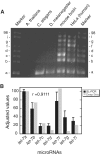
let-7 signatures in cellular, tissue and whole -organism samples as revealed by SL-PCR. (A) Total RNA preparations from various biological sources were subjected to SL-PCR in the presence of probes for human let-7 miRNAs and hsa-miR-98. (B) Comparison of let-7 signatures of HeLa cells derived from SL-PCR (black bars) and deep sequencing results (grey bars) with miRNA levels from both techniques given as values relative to 100 for let-7a. Values obtained from SL-PCR are mean ± SD of three independent experiments.
Figure 5.
Figure 5.
let-7 signatures in stem cells and during differentiation to neurons. (A) let-7 signatures of human embryonic stem cell (hESC), human mesenchymal stem cells (hMSC) and unrestricted somatic stem cells (USSC). ‘U6’ stands for human U6 snRNA (B) let-7 signatures during differentiation of hESC to neurons, driven by retinoic acid treatment in 5 days.
Figure 6.
Figure 6.
let-7 signatures in young and aged murine brain and bone marrow. let-7 miRNAs levels from tissues of five young (black bars) or five old (grey bars) mice were quantified by SL-PCR and normalized using expression levels of U6 snRNA. The data represent the mean ± SD of four repeats, *P < 0.05 and **P < 0.01. Two-tailed t-test was used to examine statistical significance.

Similar articles

Cited by

References

    1. Molnar A, Schwach F, Studholme DJ, Thuenemann EC, Baulcombe DC. miRNAs control gene expression in the single-cell alga Chlamydomonas reinhardtii. Nature. 2007;447:1126–1129. - PubMed
    1. Chaudhuri K, Chatterjee R. MicroRNA detection and target prediction: integration of computational and experimental approaches. DNA Cell. Biol. 2007;26:321–337. - PubMed
    1. Yoon S, De Micheli G. Computational identification of microRNAs and their targets. Birth Defects Res. C Embryo Today. 2006;78:118–128. - PubMed
    1. Windsor AJ, Mitchell-Olds T. Comparative genomics as a tool for gene discovery. Curr. Opin. Biotechnol. 2006;17:161–167. - PubMed
    1. Chen X. MicroRNA biogenesis and function in plants. FEBS Lett. 2005;579:5923–5931. - PMC - PubMed

Publication types