. 2011 May 1;589(Pt 9):2229-43.
doi: 10.1113/jphysiol.2010.203463. Epub 2011 Feb 28.

The Cav3.2 T-type calcium channel regulates temporal coding in mouse mechanoreceptors

Affiliations

The Cav3.2 T-type calcium channel regulates temporal coding in mouse mechanoreceptors

Rui Wang et al. J Physiol. .

Abstract

In mammals there are three types of low-voltage-activated (LVA) calcium channels,Cav3.1, Cav3.2 and Cav3.3, which all give rise to T-type Ca2+currents. T-type Ca2+currents have long been known to be highly enriched in a sub-population of medium-sized sensory neurones in the dorsal root ganglia (DRG). However, the identity of the T-type-rich sensory neurones has remained controversial and the precise physiological role of the Cav3.2 calcium channel in these sensory neurones has not been directly addressed. Here we show, using Cav3.2−/− mutant mice,that these channels are essential for the normal temporal coding of moving stimuli by specialized skin mechanoreceptors called D-hair receptors.We show that D-hair receptors from Cav3.2−/− fire approximately 50% fewer spikes in response to ramp-and-hold displacement stimuli compared to wild type receptors. The reduced sensitivity of D-hair receptors in Cav3.2−/− mice is chiefly due to an increase in the mechanical threshold and a substantial temporal delay in the onset of high-frequency firing to moving stimuli.We examined the receptive properties of other cutaneous mechano receptors and Aδ- and C-fibre nociceptors in Cav3.2−/− mice, but found no alteration in their mechanosensitivity compared to Cav3.2+/+mice. However, C-fibre nociceptors recorded in Cav3.2−/− mutant mice displayed a small but statistically significant reduction in their spiking rate during noxious heat ramps when compared to C-fibres in control mice. The T-type calcium channel Cav3.2 is thus not only a highly specific marker of D-hair receptors but is also required to maintain their high sensitivity and above all to ensure ultra rapid temporal detection of skin movement.


Figures
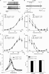
Figure 1
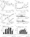
Figure 1. Stimulus response function and mechanical latencies of Aδ-mechanonociceptors in Cav3.2+/+ and Cav3.2−/− mice
A, original records for Aδ-mechanonociceptor (AM) responses to displacements of 48 μm in Cav3.2+/+ and Cav3.2−/− mice. B, an example calculation of mechanical latency for a typical C-fibre in Cav3.2−/− mice. C and E show the mean stimulus responses for A-fibre and C-fibres for the entire ramp-and-hold stimulation for the ascending displacement series. D and F, the mechanical latency of AMs and C-fibres plotted against displacement amplitude. G shows the mean firing rate of C-MH fibres during a standard ramp and hold stimulus and H, the mean temperature at which the first or second spike occured during the ramp for both genotypes.
Figure 2
Figure 2. Mechanosensitivity of D-hair receptors in Cav3.2+/+ and Cav3.2−/− mice
A, phasic response function (spike frequency during the movement of the stimuli) of D-hair receptors to a constant-ramp velocity (1432 μm s−1) stimulus with increasing displacements amplitude. B, mechanical latencies of D-hair receptors in response to increasing displacement amplitude. C, the velocity response function for D-hair receptors to a constant displacement stimulus of 96 μm with a range of ramp velocities from 6–3000 μm s−1. The mean firing frequencies are shown only for those units that fired a spike during the ramp. The percentage of responding D-hairs at the corresponding velocity step is shown in the inset. D, example trace of velocity responses of D-hairs at 356 μm s−1 in Cav3.2+/+ and Cav3.2−/− mice. E and F, detailed analysis of phasic firing frequency at velocities of 356 μm s−1 and 1432 μm s−1.
Figure 3
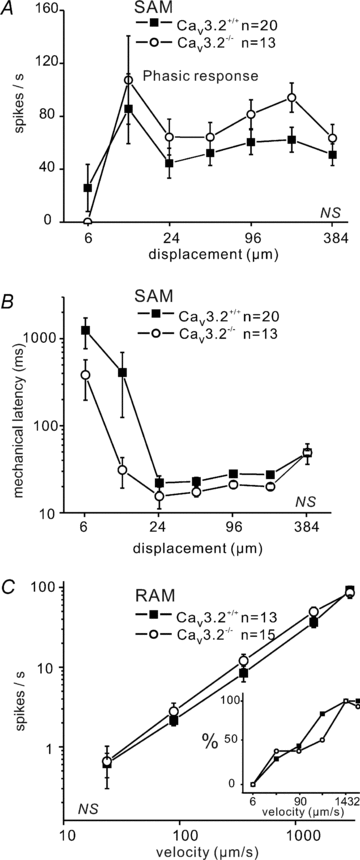
Figure 3. Mechanosensitivity of Aβ-fibres
A, phasic response function (spike frequency during the movement of the stimuli) of SAM at constant-ramp velocity (1432 μm s−1) to increasing displacements. B, mechanical latency of SAMs in response to increasing displacement amplitude. C, velocity response function for RAMs at constant displacement of 96 μm with changing velocities from 6 to 3000 μm s−1. The mean firing frequencies are shown only for those units that fired a spike during the ramp. The percentage of responding RAM at the corresponding velocity step is shown in the inset.
Figure 4
Figure 4. Mechanosensitive currents in DRG neurons
A, top, bright field image of a cultured single mechanoreceptor in the whole-cell recording configuration (RE, recording electrode) with the mechanical stimulator (MS) poised to stimulate one of the neurites. Bottom, sample trace of an RA-mechanosensitive current obtained by neurite stimulation in the presence of TTX (1 μm). B, the mean latency of RA-mechanosensitive currents (left) and mean peak current amplitude (right) for RA-mechanosensitive currents in neurones recorded from Cav3.2−/− and Cav3.2+/+ mice. Number of recorded neurones is indicated at bottom of each bar.
Figure 5
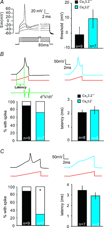
Figure 5. Threshold current for somal AP initiation
A, left panel shows stimulation protocol used to determine electrical AP threshold in mechanoreceptors (step pulses were used, starting from 500 pA with increments of 500 pA). Right panel: the mean membrane potentials at which APs are generated in Cav3.2−/− and Cav3.2+/+ mice. B, example traces of APs evoked by 5 ms current ramp injection in mechanoreceptors from both genotypes Cav3.2−/− and Cav3.2+/+. Bars on the left show the percentage of mechanoreceptors firing APs in response to 5 ms current ramp injection. Electrical latency is measured from the beginning of the current ramp to the point where the second derivative of the voltage trace reached the maximum. Mean electrical latencies are summarized in the right bar graph. C, example traces of APs evoked by 10 ms current ramp injection in mechanoreceptors from both Cav3.2−/− and Cav3.2+/+ mice. Bars on the left show the percentage of mechanoreceptors firing APs in response to 10 ms current ramp injection. Mean electrical latencies are summarized on the right bar graph. Number of recorded neurones is indicated at bottom of each bar.
Figure 6
Figure 6. A schematic diagram showing a model for AP generation in D-hair receptors in response to increasing ramp stimuli in Cav3.2+/+ and Cav3.2−/− mice
Moving stimuli evoke inward currents (row 1, from bottom upwards), reflecting the activation of RA-mechanosensitive currents (row 2). The receptor, or generator, potential (green) caused by the inward current reaches the threshold for activation of the LVA Ca2+ channel very rapidly (dotted black line) in Cav3.2+/+ mice but not in Cav3.2−/− mice (row 3). The summation of receptor potential and transient Ca2+ currents boosts the membrane depolarization to a level sufficient for regenerative APs very quickly in Cav3.2+/+ mice (pink dotted line). In contrast, in the absence of Cav3.2 channels the same receptor potential produces a much slower depolarization that delays the initial firing (row 4). As a result, APs are generated with a much shorter latency in Cav3.2+/+ than in Cav3.2−/− mice (row 5).

Comment in

Similar articles

Cited by

References

    1. Airaksinen MS, Koltzenburg M, Lewin GR, Masu Y, Helbig C, Wolf E, Brem G, et al. Specific subtypes of cutaneous mechanoreceptors require neurotrophin-3 following peripheral target innervation. Neuron. 1996;16:287–295. - PubMed
    1. Aptel H, Hilaire C, Pieraut S, Boukhaddaoui H, Mallie S, Valmier J, Scamps F. The Cav3.2/α1H T-type Ca2+ current is a molecular determinant of excitatory effects of GABA in adult sensory neurons. Mol Cell Neurosci. 2007;36:293–303. - PubMed
    1. Bal T, McCormick DA. Mechanisms of oscillatory activity in guinea-pig nucleus reticularis thalami in vitro: a mammalian pacemaker. J Physiol. 1993;468:669–691. - PMC - PubMed
    1. Barton ME, Eberle EL, Shannon HE. The antihyperalgesic effects of the T-type calcium channel blockers ethosuximide, trimethadione, and mibefradil. Eur J Pharmacol. 2005;521:79–85. - PubMed
    1. Bilici D, Akpinar E, Gursan N, Dengiz GO, Bilici S, Altas S. Protective effect of T-type calcium channel blocker in histamine-induced paw inflammation in rat. Pharmacol Res. 2001;44:527–531. - PubMed

Publication types

MeSH terms

Substances

LinkOut - more resources