. 2011 Jun 1;589(Pt 11):2755-66.
doi: 10.1113/jphysiol.2011.207282. Epub 2011 Apr 11.

Adenosine A2A receptor induces protein kinase A-dependent functional modulation of human (alpha)3(beta)4 nicotinic receptor

Affiliations

Adenosine A2A receptor induces protein kinase A-dependent functional modulation of human (alpha)3(beta)4 nicotinic receptor

Silvia Di Angelantonio et al. J Physiol. .

Abstract

Adenosine modulates the function of nicotinic ACh receptors (nAChRs) in a variety of preparations, possibly through pathways involving protein kinase A (PKA), but these phenomena have not yet been investigated in detail. In this work we studied, using the patch clamp technique, the functional modulation of recombinant human α3β4 nAChR by the A2A adenosine receptor, co-expressed in HEK cells. Tonic activation of A2A receptor slowed current decay during prolonged applications of nicotine and accelerated receptor recovery from desensitization. Together, these changes resulted into a more sustained current response upon multiple nicotine or ACh applications. These findings were confirmed in cultured mouse superior cervical ganglion neurones, which express nAChR containing the α3 subunit together with β2 and/or β4 and A2A receptor. Expression of the A2A receptor in HEK cells also increased the apparent potency of nAChR for nicotine, further supporting a general A2A-induced gain of function for nAChR. These effects were dependent on PKA since the direct activation of PKA mimicked, and its inhibition prevented almost completely, the effects of the A2A receptor. Mutations of R385 and S388 in the cytoplasmic loop of the α3 subunit abolished the functional modulation of nAChR induced by activation of A2A receptor, PKA and other Ser/Thr kinases, suggesting that this region constitutes a putative consensus site for these kinases. These data provide conclusive evidence that activation of the A2A receptor determines functional changes


Figures
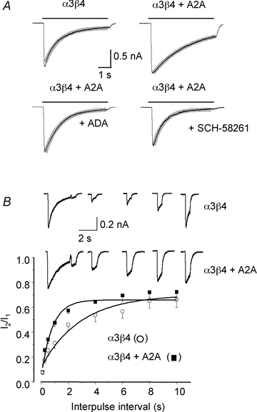
Figure 1
Figure 1. Expression of A2A receptor modulates desensitization of α3β4 nAChR
A, typical whole-cell currents evoked by nicotine (180 μm, −50 mV) in different cells expressing α3β4 nAChR plus A2A receptor, if indicated. Where indicated, adenosine deaminase (ADA, 1 U ml−1) and the A2A receptor specific blocker SCH58261 (30 nm) were present throughout the recording. Grey lines represent best fitting exponential curves of the decay phase. B, top, typical currents recorded in cells expressing α3β4 nAChR alone or with A2A receptor when a second nicotine pulse (180 μm, −50 mV) was administered at variable times after a desensitizing 5 s application. Bottom, averaged peak amplitude of the second response/first response (I2/I1) (± SEM) in cells expressing α3β4 nAChR alone (open symbols) or plus A2A receptor (filled symbols) (n = 13 and 10, respectively). Notice the slowed current decay and accelerated nAChR recovery from desensitization in cells co-expressing A2A receptor.
Figure 2
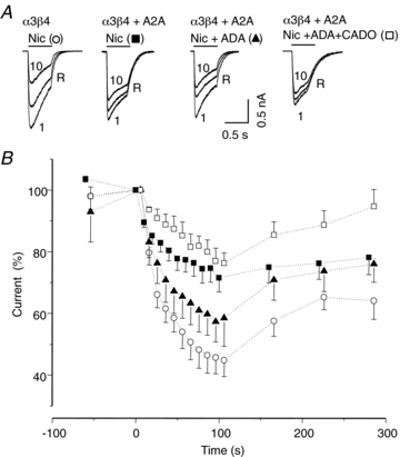
Figure 2. Current reduction upon repetitive nicotine applications is limited by A2A receptor
A, superimposed responses to nicotine (180 μm, −50 mV) in 4 different cells expressing α3β4 nAChR alone or plus A2A receptor, in the presence of ADA, or ADA plus CADO, as indicated. Currents represent the first (1) and tenth (10) response to nicotine applications at 10 s interval, and the response after 60 s wash (R). B, averaged responses (± SEM) obtained in 6 to 29 cells for each condition. In each cell, peak amplitude of subsequent responses was expressed as a percentage of the first response. ADA (1 U ml−1) or ADA plus CADO (10 nm) were present throughout the recording.
Figure 3
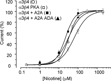
Figure 3. Nicotine concentration – current response curve
Data points were obtained in 9–21 cells transfected with α3β4 nAChR alone (open symbols) or plus A2A (filled symbols), with the indicated treatments. Error bars represent SEM, and continuous lines best fitting Hill curves, with parameters given in the text.
Figure 4
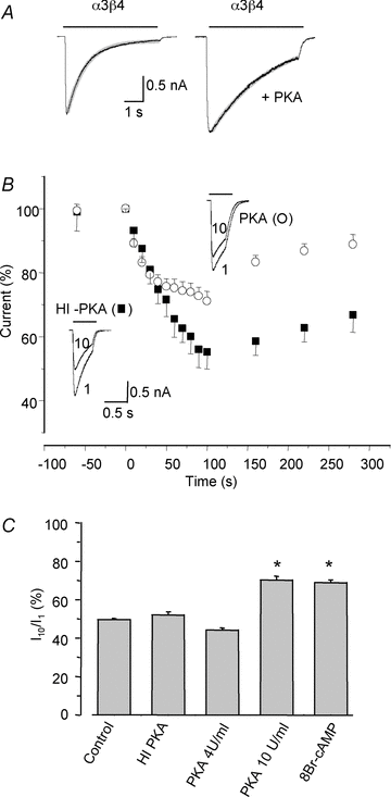
Figure 4. Protein kinase A limits current reduction upon repetitive nicotine applications to α3β4 nAChR
A, typical whole-cell currents evoked by nicotine (180 μm, −50 mV) in different cells expressing α3β4 nAChR, using control intracellular solution or intracellular solution plus PKA catalytic subunit (10 U ml−1). Grey lines represent best fitting exponential curves of the decay phase. B, averaged responses (± SEM) to repetitive nicotine applications obtained in cells dialysed with PKA catalytic subunit (10 U ml−1, filled symbols; n = 19) or the same amount of heat-inactivated (HI) enzyme (open symbols; n = 6). Values are expressed as a percentage of the peak amplitude of the first response. Insets, superimposed first (1) and tenth (10) responses to nicotine (180 μm for 0.5 s every 10 s) in cells representative of each condition. C, bar chart summarizing the ratio I10/I1, obtained from experiments as in B, averaged in 7–29 cells expressing α3β4 nAChR and treated as indicated. Holding potential, −50 mV in all recordings. *Significantly different from control value (ANOVA, P < 0.01). Notice the significant increase of the ratio I10/I1 induced by PKA.
Figure 5
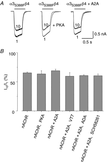
Figure 5. Mutations of α3 subunit prevent PKA effect
A, superimposed traces representing the first (1) and tenth (10) response to repetitive nicotine applications (60 μm, −50 mV) at 10 s interval in 3 different cells expressing α3S388Fβ4 nAChR alone (control or dialysed with PKA catalytic subunit 10 U ml−1) or plus A2A receptor, as indicated. B, bar chart summarizing the ratio I10/I1 averaged in 7–18 cells expressing α3S388Fβ4 nAChR and treated as indicated. Notice that no value was different from control.
Figure 6
Figure 6. Effect of other protein kinases on current reduction upon repetitive nicotine applications
A, top, superimposed traces representing the first (1) and tenth (10) response to nicotine applications (180 μm, −50 mV) repeated at 10 s interval, in cells expressing α3β4 nAChR treated with PMA to activate PKC or dialysed with CaMK II catalytic subunit (1 U ml−1). Bottom, bar chart summarizing the mean value of I10/I1 in 7 or 9 cells treated as above or in the absence of treatment (control, 9 cells). *Significantly different from control value (ANOVA, P < 0.01). B and C, as in A in cells expressing α3S388Fβ4 nAChR (5 to 8 cells) or α3R385Hβ4 nAChR (5 to 9 cells), respectively. Nicotine concentration, 60 μm. Notice that both mutations impair the effect of kinase activation.
Figure 7
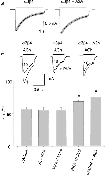
Figure 7. Current reduction upon repetitive ACh applications is limited by PKA and A2A receptor
A, typical whole-cell currents evoked by ACh (250 μm, −50 mV) in different cells expressing α3β4 nAChR alone or plus A2A receptor. Grey lines represent best fitting exponential curves of the decay phase. B, top, superimposed traces represent the first (1) and tenth (10) response to repetitive ACh applications (250 μm, −50 mV) at 10 s interval in 3 different cells expressing α3β4 nAChR alone (control or dialysed with PKA catalytic subunit 10 U ml−1) or plus A2A receptor, as indicated. Bottom, bar chart summarizing the mean value of I10/I1in 15–38 cells treated as indicated. *Significantly different from control value (ANOVA, P < 0.01).
Figure 8
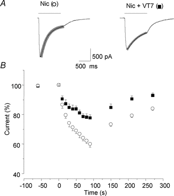
Figure 8. Activation of A2A receptor reduces decay of INic in cultured mouse neurons
A, typical whole-cell currents evoked by nicotine (100 μm, −70 mV for 1 s) alone (open symbols) or plus the A2A receptor agonist VT7 (10 nm, pre-applied for 3 min; filled symbols), in the same sympathetic neuron. Grey lines represent best fitting exponential curves of the decay phase. B, averaged responses (± SEM) obtained in 6 cells upon repetitive nicotine applications, at brief (10 s) and long (60 s) intervals under control conditions and in the presence of 10 nm VT7. In each cell, peak amplitude of subsequent responses was expressed as a percentage of the first response.

Similar articles

Cited by

References

    1. Caratsch CG, Grassi F, Eusebi F. Functional regulation of nicotinic acetylcholine receptor channels in muscle. Ion Channels. 1992;3:177–206. - PubMed
    1. Correia-de-Sá P, Ribeiro JA. Tonic adenosine A2A receptor activation modulates nicotinic autoreceptor function at the rat neuromuscular junction. Eur J Pharmacol. 1994;271:349–355. - PubMed
    1. Dani JA. Overview of nicotinic receptors and their roles in the central nervous system. Biol Psychiatry. 2001;49:166–174. - PubMed
    1. Di Angelantonio S, De Stefano ME, Piccioni A, Lombardi L, Gotti C, Paggi P. Lack of dystrophin functionally affectsα3β2/β4-nicotinic acethylcholine receptors in sympathetic neurons of dystrophic mdx mice. Neurobiol Dis. 2011;41:528–537. - PubMed
    1. Downing JE, Role LW. Activators of protein kinase C enhance acetylcholine receptor desensitization in sympathetic ganglion neurons. Proc Natl Acad Sci U S A. 1987;84:7739–7743. - PMC - PubMed

Publication types

MeSH terms

Substances

LinkOut - more resources