. 2011 Jun 15;589(Pt 12):2927-34.
doi: 10.1113/jphysiol.2011.206672. Epub 2011 Apr 11.

Lymphatic anatomy and biomechanics

Affiliations

Lymphatic anatomy and biomechanics

Daniela Negrini et al. J Physiol. .

Abstract

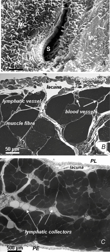
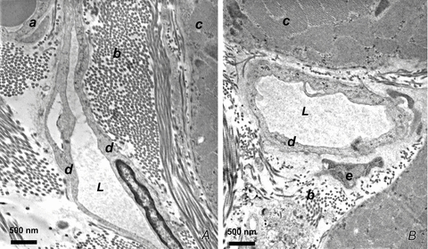
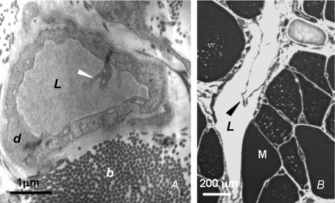
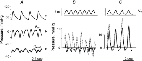
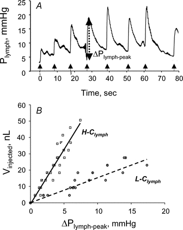
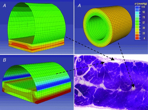
Lymph formation is driven by hydraulic pressure gradients developing between the interstitial tissue and the lumen of initial lymphatics. While in vessels equipped with lymphatic smooth muscle cells these gradients are determined by well-synchronized spontaneous contractions of vessel segments, initial lymphatics devoid of smooth muscles rely on tissue motion to form lymph and propel it along the network. Lymphatics supplying highly moving tissues, such as skeletal muscle, diaphragm or thoracic tissues, undergo cyclic compression and expansion of their lumen imposed by local stresses arising in the tissue as a consequence of cardiac and respiratory activities. Active muscle contraction and not passive tissue displacement is required to support an efficient lymphatic drainage, as suggested by the fact that the respiratory activity promotes lymph formation during spontaneous, but not mechanical ventilation. The mechanical properties of the lymphatic wall and of the surrounding tissue also play an important role in lymphatic function. Modelling of stress distribution in the lymphatic wall suggests that compliant vessels behave as reservoirs accommodating absorbed interstitial fluid, while lymphatics with stiffer walls, taking advantage of a more efficient transmission of tissue stresses to the lymphatic lumen, propel fluid through the lumen of the lymphatic circuit.


Figures

Figure 1
Figure 1. Arrangement of the diaphragmatic lymphatic network
A, scanning electron photomicrograph of an elliptical lymphatic stoma on the tendinous pleural surface of the rat diaphragm. The stoma (S), delimited by a net border, opens at the confluence between adjacent mesothelial cells (MC) characterized by a mesh of microvilli protruding from the cell surface. B, semi-thin cross-section of rat pleural hemi-diaphragm stained with crystal violet and basic fuchsin showing the arrangement of the diaphragmatic lymphatic network originating from the pleural mesothelial surface. Lymphatic submesothelial lacunae located within the interstitial space beneath the mesothelial layer are in continuity with transverse lymphatic vessels (arrow) located in the deep interstitial space and running perpendicularly to the lacunae and through the diaphragmatic skeletal muscular fibres. C, semi-thin cross-section of rat entire diaphragm stained with crystal violet and basic fuchsin, showing both the pleural (PL) and the peritoneal (PE) mesothelial surfaces. Transverse lymphatic vessels departing from the pleural and peritoneal submesothelial lacunae empty into central lymphatic collectors, located in the deep interstitial space, which receive the lymph collected from both surfaces of the diaphragm.
Figure 2
Figure 2. Anatomical proximity of initial lymphatic lumen and interstitial fibrillar structures
A and B, transmission electron microphotographs of thin sections of rat diaphragm. Initial lymphatics run through connective tissue composed of loose collagen fibres organized in bundles (b) adjacent to the skeletal muscular fibres (c). These vessels are characterized by typical ultrastructural features: absence of smooth muscle fibres, thin endothelial wall, discontinuous basal lamina, tight junctions or overlapping contacts and anchoring filaments linking the endothelial cells to the adjacent collagen fibres and to muscle fibre sarcolemma. L, lymphatic lumen; a, vascular capillary endothelium; b, collagen bundles; c, skeletal muscle fibres; d, lymphatic endothelium; e, fibroblast.
Figure 3
Figure 3. Lymphatic unidirectional valves
A, transmission electron microphotographs showing the primary unidirectional valves (white arrowhead) in the wall of diaphragmatic initial lymphatics located in the pleural submesothelial interstitium. The valves are formed at the junction between overlapping cytoplasmic extensions of two adjacent lymphatic endothelial cells (d) (reproduced from Grimaldi et al. 2006). B, semi-thin sections of transverse lymphatic ducts, showing a unidirectional intraluminar, or secondary, valve formed by two leaflets (arrowhead) attached at opposite sides of the lymph channel wall. L, lymphatic lumen; b, collagen bundles; M, skeletal muscle fibres; d, lymphatic endothelium (reproduced from Grimaldi et al. 2006).
Figure 4
Figure 4. Effect of cardiac and respiratory activity on interstitial (Pint) and lymphatic (Plymph) fluid pressure
A, cardiogenic oscillations of diaphragmatic interstitial (b, Pint) and intraluminar lymphatic pressure (c, Plymph), measured through the micropuncture technique in anaesthetized rats under neuromuscular blockade. Both diaphragmatic Pint and Plymph oscillate almost in phase with arterial systemic pressure (a), shifting from a minimum to a maximum value during cardiogenic oscillations (modified from Negrini et al. 2004). B and C, simultaneous recording of respiratory tidal volume (VT, top panel), lymphatic (Plymph; continuous lines) and interstitial (Pint; dashed lines) pressures obtained in intercostal lymphatics of supine anaesthetized rats during spontaneous breathing (B) or mechanical ventilation at similar VT and at zero alveolar end-expiratory pressure (C) (modified from Moriondo et al. 2005).
Figure 5
Figure 5. Pressure–volume behaviour of submesothelial diaphragmatic lymphatics
A, time course of Plymph from the pre-injection value at t = 0 and during sequential injections of 4.6 nl of lissamine green saline solution. At each injection Plymph sharply increases by ΔPlymph-peak to attain a peak value followed by a slower Plymph decay to pre-injection values (modified from Moriondo et al. 2010). B, relationships obtained by pooling data from several measurements in different diaphragmatic submesothelial vessels, where the cumulative injected volume, Vinjected, is plotted as a function of ΔPlymph-peak. The data points appear distributed in two distinct populations characterized by a significantly (P < 0.05) different Clymph amounting to 6.7 nl mmHg−1 (high compliance, H-Clymph) and 1.5 nl mmHg−1 (low compliance, L-Clymph), respectively (modified from Moriondo et al. 2010).
Figure 6
Figure 6. Three-dimensional modelling of the lymphatic vessel wall
Stress distribution maps obtained through finite element modelling of diaphragmatic initial lymphatics located as indicated on the microphotograph of the transverse section of the diaphragm, panel D. A, immediately beneath the mesothelium: the ellipsoidal superficial vessel is limited mostly by a thin wall of lymphatic endothelium plus pleural mesothelium and lies on a diaphragmatic muscular/tendinous support. B, deeper in the submesothelial tissue: this intermediate vessel is only partially delimited by a thin wall, most of the lateral surface being surrounded by the muscular/tendinous tissue. C, among the diaphragmatic muscular/tendinous fibres surrounded by an isotropic tissue and with a circular cross sectional area (modified from Moriondo et al. 2010). The circumferential stress (σ) distribution is identified by colours on a scale from red (low stress) to blue (high stress) as indicated by the colour scale in C. Vessel tensile stress was higher in submesothelial superficial (A) and intermediate (B) vessels which underwent the greatest deformation. In deeper circular vessels (C) surrounded by stiffer tissue, wall tension was lower and homogeneously distributed over the entire surface.

Similar articles

Cited by

References

    1. Aukland K, Reed R. Interstitial–lymphatic mechanisms in the control of extracellular fluid volume. Physiol Rev. 1993;73:1–78. - PubMed
    1. Benoit JN, Zawieja DC, Goodman AH, Granger HJ. Characterization of intact mesenteric lymphatic pump and its responsiveness to acute edemagenic stress. Am J Physiol Heart Circ Physiol. 1989;257:H2059–H2069. - PubMed
    1. Burgeson RE, Lunstrum GP, Rokosova B, Rimberg CS, Rosenbaum LM, Keene DR. The structure and function of type VII collagen. Ann N Y Acad Sci. 1990;580:32–43. - PubMed
    1. Casley-Smith JR, Florey HW. The structure of normal small lymphatics. Q J Exp Physiol Cogn Med Sci. 1961;46:101–106. - PubMed
    1. Castenholz A. Structural and functional properties of initial lymphatics in the rat tongue: scanning electron microscopic findings. Lymphology. 1987;20:112–125. - PubMed

LinkOut - more resources