. 2011 May 1;124(Pt 9):1553-63.
doi: 10.1242/jcs.080580. Epub 2011 Apr 12.

Canonical Wnt signaling promotes the proliferation and neurogenesis of peripheral olfactory stem cells during postnatal development and adult regeneration

Affiliations

Canonical Wnt signaling promotes the proliferation and neurogenesis of peripheral olfactory stem cells during postnatal development and adult regeneration

Ya-Zhou Wang et al. J Cell Sci. .

Abstract

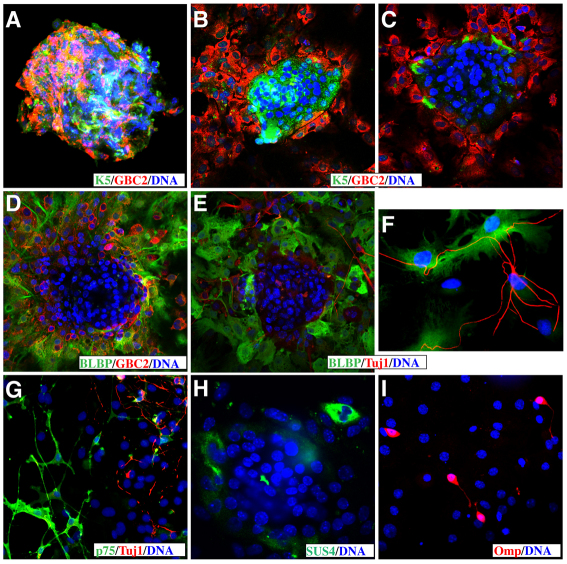
The mammalian olfactory epithelium (OE) has a unique stem cell or progenitor niche, which is responsible for the constant peripheral neurogenesis throughout the lifespan of the animal. However, neither the signals that regulate the behavior of these cells nor the lineage properties of the OE stem cells are well understood. Multiple Wnt signaling components exhibit dynamic expression patterns in the developing OE. We generated Wnt signaling reporter TOPeGFP transgenic mice and found TOPeGFP activation predominantly in proliferating Sox2(+) OE basal cells during early postnatal development. FACS-isolated TOPeGFP(+) OE basal cells are required, but are not sufficient, for formation of spheres. Wnt3a significantly promotes the proliferation of the Sox2(+) OE sphere cells. Wnt-stimulated OE sphere cells maintain their multipotency and can differentiate into most types of neuronal and non-neuronal epithelial cells. Also, Wnt activators shift the production of differentiated cells toward olfactory sensory neurons. Moreover, TOPeGFP(+) cells are robustly increased in the adult OE after injury. In vivo administration of Wnt modulators significantly alters the regeneration potential. This study demonstrates the role of the canonical Wnt signaling pathway in the regulation of OE stem cells or progenitors during development and regeneration.


Figures

Fig. 1.
Fig. 1.
The expression of TOPeGFP reporter transgene and Wnt signaling components in mouse olfactory epithelium (OE) at P5. (A) Schematic illustration of the TOPeGFP transgenic construct. (B) eGFP+ cells (arrows) extend throughout the OE in a representative coronal section of the TOPeGFP transgenic mice. The dashed rectangle indicates the septal region of the OE (roughly equivalent to region shown in D–L). (C) Real-time RT-PCR for representative Wnt signaling molecules Axin2, Tcf7l2, Fzd9 and Wnt3a as well as GFP in GFP+ and GFP cells isolated by FACS from P5 OE tissue of TOPeGFP mice. The relative mRNA level was normalized to Gapdh. *P<0.05; **P<0.01. (DH) Colocalization of TOPeGFP (green) with mRNA signals (purple or brown) of Axin2, Tcf7l2 and Fzd9 in the basal cells (arrows and arrowheads) of P5 OE. The fluorescence microscopy images of eGFP were taken directly (without immunohistochemistry) before the in situ hybridization and inverted to bright fields in F–H. (IL) In situ hybridization demonstrates dynamic expression patterns of representative canonical ligands Wnt3a, Wnt2 and Wnt7b, and an inhibitory ligand Sfrp1 in the P5 OE. Asterisks in K and L indicate the pattern of gene expression in sustentacular cells (Sus). The arrows in L indicate Sfrp1 expression in the lamina propria (LP). OSN, olfactory sensory neurons. Scale bars: 200 μm (B), 50 μm (D–L).
Fig. 2.
Fig. 2.
Double immunolabeling of TOPeGFP with markers for OE basal cells, proliferation and immature or mature OSNs. (A) eGFP and Sox2 double immunolabeling. Arrows indicate the colocalization of eGFP and Sox2 (a stem cell marker) immunoreactivity in the HBC layer. Arrowheads indicate the double-labeled cells in the GBC layer. Asterisk indicates the Sox2+/GFP sustentacular cell layer. (B) eGFP and K5 double immunolabeling. Arrows indicate the colocalization of eGFP and K5 (a HBC marker) immunoreactivity in the HBC layer. Arrowheads indicate a basal cell strongly labeled for eGFP and weak or no K5 immunolabeling. (C) Double immunolabeling for eGFP and acute BrdU (2 hour incorporation) in P5 OE. Arrows indicate many double-immunolabeled basal cells. (D) Double immunolabeling for eGFP and BrdU (8 hour incorporation from four injections) in P5 OE. Arrows indicate more double-immunolabeled cells in the HBC layer. Arrowheads indicate many double-immunolabeled cells in the GBC and OSN layers. (E) Double immunolabeling for eGFP and Dcx (an immature neuronal marker). Arrowheads indicate some double-immunolabeled cells in the GBC and immature OSN layers. (F) Most Omp+ mature OSNs are GFP. (G) Percentage of OE basal cells or OSNs (red bars) in TOPeGFP+ cells and percentage of Wnt-responsive cells (green bars) in various OE cell groups immunolabeled with different markers. Cell nuclei were counterstained by DAPI (blue). Data are means ± s.e.m. Scale bars: 50 μm.
Fig. 3.
Fig. 3.
Wnt signaling promotes primary and secondary OE sphere formation. (A) RT-PCR analysis of representative Wnt signaling components in primary OE spheres. The negative controls omitted the reverse transcription step. (B) TOPeGFP activation in OE spheres was enhanced by the Wnt activator (Gsk3 inhibitor) BIO and suppressed by the Wnt inhibitory ligand Dkk1 as measured by direct fluorescence microscopy. (C) Activators of Wnt signaling (Wnt3a, LiCl, and BIO) increased, whereas Dkk1 inhibited primary OE sphere formation. (D) Wnt3a, LiCl and BIO promoted secondary sphere formation, whereas the inhibitor Dkk1 repressed it. (E) The GFP gate in FL1 was set with reference to the distribution of unlabeled cells from the wild-type mice for FACS; about 33.69% of the gated cells were GFP+ in TOPeGFP mice. Cy5 channel (Y axis) was used for compensation. (F) BIO increased OE sphere formation of both unsorted and GFP+ sorted cells. The frequency of sphere formation from sorted GFP+ cells was significantly lower than that obtained from the unsorted OE cells. Very few OE spheres were found in the sorted GFP OE cells. *P<0.05; **P<0.01.
Fig. 4.
Fig. 4.
Effects of Wnt3a stimulation on the proliferation and apoptosis of dissociated OE sphere cells. (A,B,G) The percentage of Sox2+, BrdU+ or BrdU+Sox2+ OE sphere cells was significantly increased by Wnt3a stimulation compared with PBS control culture. (CG) No significant differences of the percentage of Caspase3+ or TUNEL+ apoptotic cells existed between the Wnt3a treated and control cultures. P<0.05 is considered statistically significant.
Fig. 5.
Fig. 5.
Multipotency of OE spheres cultured with Wnt activators. (A) An undifferentiated adherent OE sphere co-immunolabeled with K5 (for HBCs) and GBC2 (for the GBC lineage). (BI) Differentiated adherent sphere cells immunolabeled with lineage-specific marker antibodies including K5 and GBC2 (B,C), BLBP (for olfactory glial lineage) in combination with GBC2 (D), BLBP in combination with Tuj1 (olfactory neuronal lineage) (E,F), p75 (OECs) double immunolabeling with Tuj1 (G), SUS4 (for sustentacular cells) (H) and Omp (for mature OSNs) (I). Cell nuclei were counterstained with DAPI.
Fig. 6.
Fig. 6.
Effects of Wnt stimulation on differentiation from dissociated OE sphere cells. (A,B) The percentage of Tuj1+ OSNs was significantly increased by treatment with Wnt3a, LiCl and BIO compared with the controls. (C,D) The percentage of BLBP+ glial lineage cells was decreased by treatment with Wnt signaling activators. *P<0.05; **P<0.01.
Fig. 7.
Fig. 7.
Dramatic changes in the TOPeGFP+ basal cells and OE morphology during forced OE regeneration in adult mice. (AA″) Relatively low activity of TOPeGFP expression (immunolabeled with brown DAB products) in the control animal injected with saline. (BD″) Robustly increased TOPeGFP expression during forced OE regeneration at 3 days (B-B″), 1 week (C-C″), and 2 weeks (D-D″) after intraperitoneal administration with methimazole. Dashed rectangles in A–D indicate respective enlarged regions in A′–D″. Asterisks in B–D indicate the degenerated OE-like tissue. Arrows in A′–D″ indicate the surface mucous layer. Red lines in A′–D′ indicate the thickness of the normal OE and the changes of the TOPeGFP+ basal layers. I, superior nasal turbinate; IIa, IIb, branches of the middle turbinate; III, VI, inferior turbinates; S, septum. Scale bars: 500 μm.
Fig. 8.
Fig. 8.
In vivo administration of small molecules of Wnt signaling modulators altered OE regeneration in adult mice. (AE) Intraperitoneal administration of a Wnt signaling inhibitor Quercetin significantly reduced the numbers of Omp+ OSNs, BrdU+ proliferating and TOPeGFP+ basal cells in the septal OE after 2 weeks of methimazole treatment. (FJ) Intraperitoneal administration of the Wnt signaling activator lithium significantly increased the numbers of Omp+ OSNs and TOPeGFP+ basal cells at 7 days and BrdU+ proliferating OE basal cells at 3 days, in the septal OE after methimazole treatment. Dashed lines indicate the basal membrane dividing OE and lamina propria. *P<0.05; **P<0.01.

Similar articles

Cited by

References

    1. Agathocleous M., Iordanova I., Willardsen M. I., Xue X. Y., Vetter M. L., Harris W. A., Moore K. B. (2009). A directional Wnt/beta-catenin-Sox2-proneural pathway regulates the transition from proliferation to differentiation in the Xenopus retina. Development 136, 3289-3299 - PMC - PubMed
    1. Bandyopadhyay U., Biswas K., Banerjee R. K. (2002). Extrathyroidal actions of antithyroid thionamides. Toxicol. Lett. 128, 117-127 - PubMed
    1. Barraud P., He X., Zhao C., Ibanez C., Raha-Chowdhury R., Caldwell M. A., Franklin R. J. (2007). Contrasting effects of basic fibroblast growth factor and epidermal growth factor on mouse neonatal olfactory mucosa cells. Eur. J. Neurosci. 26, 3345-3357 - PubMed
    1. Bergman U., Ostergren A., Gustafson A. L., Brittebo B. (2002). Differential effects of olfactory toxicants on olfactory regeneration. Arch. Toxicol. 76, 104-112 - PubMed
    1. Bergstrom U., Giovanetti A., Piras E., Brittebo E. B. (2003). Methimazole-induced damage in the olfactory mucosa: effects on ultrastructure and glutathione levels. Toxicol. Pathol. 31, 379-387 - PubMed

Publication types

MeSH terms