. 2011 May 1;124(Pt 9):1486-95.
doi: 10.1242/jcs.082834. Epub 2011 Apr 12.

Truncation of monocyte chemoattractant protein 1 by plasmin promotes blood-brain barrier disruption

Affiliations

Truncation of monocyte chemoattractant protein 1 by plasmin promotes blood-brain barrier disruption

Yao Yao et al. J Cell Sci. .

Abstract

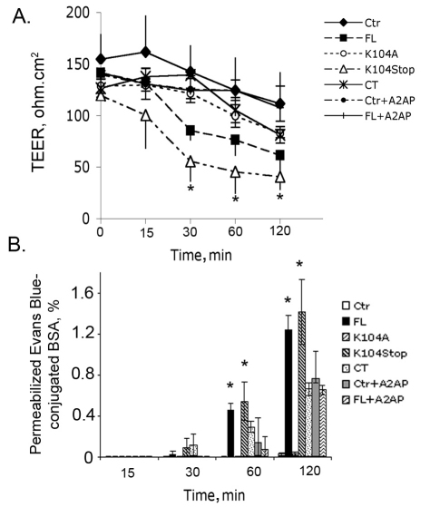
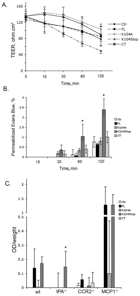
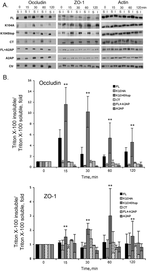
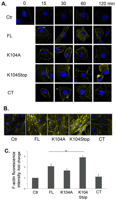
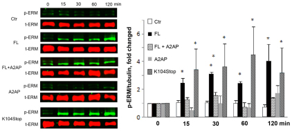
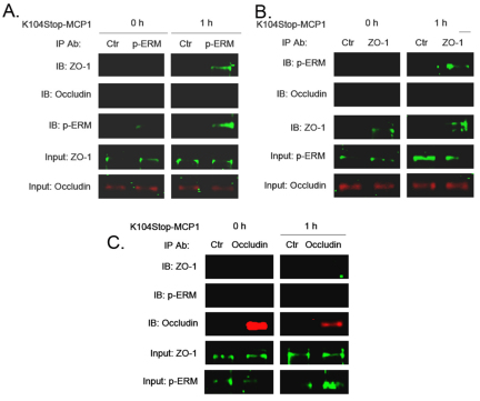
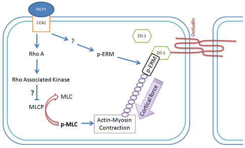
Previous studies have shown that plasmin cleaves monocyte chemoattractant protein 1 (MCP1; officially known as C-C motif chemokine 2, CCL2) at K104, and this cleavage enhances its chemotactic potency significantly. Accumulating evidence reveals that MCP1 also disrupts the integrity of the blood-brain barrier (BBB). Here, we show that K104Stop-MCP1, truncated at the K104 where plasmin would normally cleave, is more efficient than the full-length protein (FL-MCP1) in compromising the integrity of the BBB in in vitro and in vivo models. K104Stop-MCP1 increases the permeability of BBB in both wild-type mice and mice deficient for tissue plasminogen activator (tPA), which converts plasminogen into active plasmin, suggesting that plasmin-mediated truncation of MCP1 plays an important role in BBB compromise. Furthermore, we show that the mechanisms underlying MCP1-induced BBB disruption involve redistribution of tight junction proteins (occludin and ZO-1) and reorganization of the actin cytoskeleton. Finally, we show that the redistribution of ZO-1 is mediated by phosphorylation of ezrin-radixin-moesin (ERM) proteins. These findings identify plasmin as a key signaling molecule in the regulation of BBB integrity and suggest that plasmin inhibitors might be used to modulate diseases accompanied by BBB compromise.


Figures

Fig. 1.
Fig. 1.
Truncated MCP1 compromises the integrity of the HBMEC monolayer. HBMECs seeded in Transwell inserts were treated with the indicated form of MCP1 (100 nM; in the lower chamber, abluminal side) in the presence or absence of A2AP. Ctr, control (saline). (A) Changes in the transendothelial electrical resistance (TEER) over time. Results are means±s.d. (n=3). *P<0.05 (analyzed using one-way ANOVA followed by Newman–Keuls multiple comparison test) compared with results with FL-MCP1 without A2AP. (B) Chances in the permeability of Evan Blue over time. Results are means±s.d. (n=6). *P<0.05 (comparison with Ctr).
Fig. 2.
Fig. 2.
Truncated MCP1 compromises BBB integrity. (A) The indicated form of MCP1 (100 nM) was added to the lower chamber of the BMEC-astrocyte co-culture system. TEER values were determined over time in the presence of A2AP. Results are means±s.d. (n=3). Ctr, control (saline). (B) Cells were treated as in A. The leakage of Evans Blue across the co-culture system was determined spectrophotometrally after treatment with A2AP. Results are means±s.d. (n=4). *P<0.05 (analyzed using one-way ANOVA followed by Newman–Keuls multiple comparison test) compared with results with FL-MCP1. (C) Infiltration of Evans Blue across the compromised BBB into brain parenchyma was assayed 12 hours after a single injection of MCP1 (1 μg). Mice injected with saline were used as control. Results [OD (optical density; absorbance) per g of body weight] are means+s.d. (n=4). *P<0.05 (analyzed using one-way ANOVA followed by Newman–Keuls multiple comparison test) compared with results with FL-MCP1 without A2AP.
Fig. 3.
Fig. 3.
Truncated MCP1 induces the redistribution of tight junction proteins. (A) HBMECs were treated with the indicated form of MCP1 (100 nM). Ctr, control (saline); WT, wild-type. At 15, 30, 60 and 120 minutes after treatment, the cells were fixed and immunostained for occludin and ZO-1. (B) Higher magnification of cells incubated with the MCP-1 variants and stained for occludin at the 120-minute timepoint. Scale bar: 15 μm. (C) Primary mouse BMECs were exposed to the MCP1 variants (100 nM) for 2 hours. Then, the cells were fixed and immunostained for ZO-1 and occludin. Blue, DAPI staining.
Fig. 4.
Fig. 4.
Truncated MCP1 shifts TJPs from the Triton-X-100-soluble fraction to the Triton-X-100-insoluble fraction. HBMECs were exposed to the indicated form of MCP1 (100 nM) for different periods of time in the presence or absence of A2AP. Ctr, control (saline). The shift of occludin and ZO-1 from the Triton-X-100-soluble fraction (S) to the Triton-X-100-insoluble fraction (I) was analyzed by western blotting. Actin was used a loading control. (A) Representative images of western blots. (B) Quantitative data from western blots. The intensity of target bands was determined using Scion Image. The intensity of occludin and ZO-1 was normalized to that of actin and the ratios were further normalized to the zero timepoint. Results are means±s.d. (n=3). **P<0.01 (analyzed using one-way ANOVA followed by Newman–Keuls multiple comparison test) for K104Stop-MCP1 compared with FL-MCP1 at each timepoint.
Fig. 5.
Fig. 5.
Truncated MCP1 promotes reorganization of the actin cytoskeleton. (A) HBMECs were treated with saline (Ctr) or the indicated form of MCP1 (100 nM) for different times, and the actin cytoskeleton was visualized using Alexa-Fluor-647–phalloidin. (B) Primary mouse BMECs treated with the indicated form of MCP1 (100 nM) for 2 hours were immunostained to visualize the actin cytoskeleton. (C) Quantification of F-actin fluorescence intensity. Values are mean+s.d. (n=9). *P<0.05 (analyzed using Student's t-tests).
Fig. 6.
Fig. 6.
Truncated MCP1 induces phosphorylation of ERM proteins. Mouse BMECs were treated with saline (Ctr, control) or the indicated form of recombinant MCP1 proteins (100 nM) with or without A2AP over time. The phosphorylated ERM proteins (p-ERM) were determined using a phosphorylation-specific antibody for ERM proteins. The level of total ERM (t-ERM) proteins is also shown. Quantitative data of western blots are shown as mean+s.d. (n=3). *P<0.05 (compared with the zero timepoint control).
Fig. 7.
Fig. 7.
Truncated MCP1 promotes interaction between phosphorylated ERM proteins and ZO-1. Mouse BMECs were treated with K104Stop-MCP1 or saline (0 h) for 1 hour. Cell lysates were collected and used for co-immunoprecipitation experiments. (A) Immunoprecipitation (IP) with an anti-(phosphorylated ERM) antibody (p-ERM) and immunoblotting (IB) with anti-ZO-1 and anti-occludin antibodies. (B) Immunoprecipitation with an anti-ZO-1 antibody and immunoblotting with anti-(phosphorylated ERM) and anti-occludin antibodies. (C) Immunoprecipitation with an anti-occludin antibody and immunoblotting with anti-(phosphorylated ERM) and anti-ZO-1 antibodies. Anti-(mouse IgG) antibody was used as control (Ctr).
Fig. 8.
Fig. 8.
Proposed model for MCP1-induced BBB compromise. Upon MCP1 treatment, ERM proteins are phosphorylated by unknown kinases. The phosphorylated ERM proteins then bind to ZO-1 and the actin cytoskeleton. By binding to CCR2, MCP1 also activates RhoA, and Rho-associated kinases, which phosphorylate and inactivate MLCP, leading to enhanced phosphorylation of MLC and thus prolonged actin–myosin contraction. The contractile forces then pull ZO-1 away from the cell-cell border, resulting in a disrupted BBB.

References

    1. Ahdieh M., Vandenbos T., Youakim A. (2001). Lung epithelial barrier function and wound healing are decreased by IL-4 and IL-13 and enhanced by IFN-gamma. Am. J. Physiol. Cell Physiol. 281, C2029-C2038 - PubMed
    1. Alter A., Duddy M., Hebert S., Biernacki K., Prat A., Antel J. P., Yong V. W., Nuttall R. K., Pennington C. J., Edwards D. R., et al. (2003). Determinants of human B cell migration across brain endothelial cells. J. Immunol. 170, 4497-4505 - PubMed
    1. Blamire A., Anthony D., Rajagopalan B., Sibson N., Perry V., Styles P. (2000). Interleukin-1beta-induced changes in blood-brain barrier permeability, apparent diffusion coefficient, and cerebral blood volume in the rat brain: a magnetic resonance study. J. Neurosci. 20, 8153-8159 - PMC - PubMed
    1. Bretscher A., Gary R., Berryman M. (1995). Soluble ezrin purified from placenta exists as stable monomers and elongated dimers with masked C-terminal ezrin-radixin-moesin association domains. Biochemistry 34, 16830-16837 - PubMed
    1. Calabria A. R., Weidenfeller C., Jones A. R., de Vries H. E., Shusta E. V. (2006). Puromycin-purified rat brain microvascular endothelial cell cultures exhibit improved barrier properties in response to glucocorticoid induction. J. Neurochem. 97, 922-933 - PubMed

Publication types

MeSH terms