. 2011 May 1;124(Pt 9):1425-32.
doi: 10.1242/jcs.077388. Epub 2011 Apr 12.

Actomyosin-generated tension controls the molecular kinetics of focal adhesions

Affiliations

Actomyosin-generated tension controls the molecular kinetics of focal adhesions

Haguy Wolfenson et al. J Cell Sci. .

Abstract

Focal adhesions (FAs) have key roles in the interaction of cells with the extracellular matrix (ECM) and in adhesion-mediated signaling. These dynamic, multi-protein structures sense the ECM both chemically and physically, and respond to external and internal forces by changing their size and signaling activity. However, this mechanosensitivity is still poorly understood at the molecular level. Here, we present direct evidence that actomyosin contractility regulates the molecular kinetics of FAs. We show that the molecular turnover of proteins within FAs is primarily regulated by their dissociation rate constant (k(off)), which is sensitive to changes in forces applied to the FA. We measured the early changes in k(off) values for three FA proteins (vinculin, paxillin and zyxin) upon inhibition of actomyosin-generated forces using two methods - high temporal resolution FRAP and direct measurement of FA protein dissociation in permeabilized cells. When myosin II contractility was inhibited, the k(off) values for all three proteins changed rapidly, in a highly protein-specific manner: dissociation of vinculin from FAs was facilitated, whereas dissociation of paxillin and zyxin was attenuated. We hypothesize that these early kinetic changes initiate FA disassembly by affecting the molecular turnover of FAs and altering their composition.


Figures
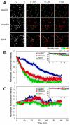
Fig. 1.
Fig. 1.
Effect of blebbistatin on FA reorganization in HeLa cells expressing YFP-tagged paxillin, vinculin or zyxin. Frames from time-lapse movies were analyzed by temporal ratio imaging. (A) Ratio between the intensity at time point 0 (first column) and time points 10, 30 and 60 minutes. Scale bar: 5 μm. Red shift denotes structures with decreased intensity; blue shift denotes an increase in intensity; green and yellow hues mark unchanged pixels. Whereas paxillin showed a delayed reaction to blebbistatin, vinculin and zyxin responded much faster (compare time points 0’:10’ and 0’:30’). (B,C) Mean relative FA fluorescence (± s.e.m., in lighter hues) over time, as derived from the time-lapse movies (n=10–15). FAs in cells treated with blebbistatin disassemble over time, resulting in very small FAs (B), whereas FAs in untreated cells remain stable over time, and their mean relative fluorescence levels fluctuate around 100% (C).
Fig. 2.
Fig. 2.
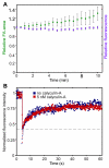
Effects of calyculin A treatment on FA area, fluorescence intensity and FRAP in HeLa cells expressing paxillin–YFP. (A) Live-cell imaging was used to monitor the relative FA area (green) and fluorescence intensity (purple) in response to calyculin A treatment. Images were taken every 30 seconds, and the resulting data were analyzed using ImageJ. During the 10 minute incubation with calyculin A, a gradual increase in FA area was noticed, without influencing paxillin–YFP intensity. Values are means ± s.e.m. (B) Typical FRAP curves of YFP-tagged paxillin before (blue) and after (red) treatment with calyculin A. Solid lines denote the best fit of a least squares regression analysis. Empty data points below the dashed line correspond to recovery by diffusion; the slower phase represents proteins that recover by exchange (Wolfenson et al., 2009b). No significant change in the exchange rate of paxillin–YFP was noted within the 10 minute interval following calyculin A treatment.
Fig. 3.
Fig. 3.
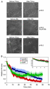
Effects of force reduction on cell contraction and FA area. (A) Phase-contrast images of HeLa cells plated on coverslips pre-coated with elastic silicon substrata. Contraction of the cells induces wrinkling of the silicon surface (Harris et al., 1980). Once the wrinkles are created by the cells, they usually persist for several hours. Top panels, HeLa cells in medium, before (left) and after (right) addition of Triton X-100 (0.1% final concentration in 20 mM HEPES buffer, pH 6.8, for 10 minutes). Virtually no change in the wrinkles is evident following permeabilization of the cells, suggesting that the cellular tension is unaltered under these conditions. Middle panels, cells before (left) and after addition of blebbistatin (Blb) to a final concentration of 50 μM (right). Note that the wrinkles vanish almost completely. Bottom panels, cells before (left) and after (right) permeabilization with Triton X-100 in 50 mM MES buffer, pH 6.2 (tension-relaxing conditions) (Metzger and Moss, 1990). The wrinkles disappear within 1–2 minutes of permeabilization at the lower pH. (B) Effect of blebbistatin on FA area in HeLa cells expressing YFP-tagged paxillin, vinculin or zyxin. Mean relative FA areas (± s.e.m., in lighter hues) over time were derived from time-lapse movies (n=10–15) of cells treated with blebbistatin. For all three proteins, the total FA area decreased essentially instantaneously following blebbistatin treatment, reaching a value of ~60% of the initial areas within 8 minutes. From that point onwards, the rates at which the area decreased differed among the three proteins: for paxillin, the FA area decreased gradually to 20% of the initial area after 60 minutes; for vinculin, the FA area continued to drop at a similar rate for an additional 4 minutes, and then gradually decreased to 10% of the initial area after 60 minutes; for zyxin, the FA area continued to drop at a similar rate for an additional 4 minutes, but unlike vinculin, it then stabilized at 40% of the initial area, within 60 minutes after treatment with blebbistatin.
Fig. 4.
Fig. 4.
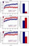
Effect of blebbistatin treatment on FRAP in FAs. Typical FRAP curves (left panels) of YFP-tagged paxillin, vinculin or zyxin, with (red) or without blebbistatin (blue). Solid lines denote the best fit of a least squares regression analysis. Average τex and koff values for each protein are shown (n=20–30). Empty data points below the dashed line correspond to recovery by diffusion; the slower phase represents proteins that recover by exchange (Wolfenson et al., 2009b). Mean ± s.e.m. (n=20–30) of the fractions undergoing exchange are shown on the right (*P<10−4; Student's t-test). For paxillin and zyxin, blebbistatin induced a decrease in the exchange rates, accompanied by lower exchanging fractions. For vinculin, blebbistatin induced the opposite effect, increasing both the exchange rate and the exchanging fraction.
Fig. 5.
Fig. 5.
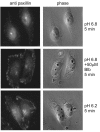
Effect of reduction of actomyosin-generated forces on paxillin dissociation from FAs. Fluorescent (left) or phase-contrast images (right) of HeLa cells plated on FN-coated coverslips. Cells were incubated (5 minutes) in either HEPES buffer, pH 6.8, supplemented with Triton X-100, with or without 1 minute pre-incubation with blebbistatin, or in MES buffer, pH 6.2, supplemented with Triton X-100. Cells were then fixed and immunolabeled for paxillin. At pH 6.8, in the absence of blebbistatin, almost no FAs were detected. However, substantial amounts of FAs were identified in cells pre-incubated with blebbistatin at pH 6.8, or incubated at pH 6.2.
Fig. 6.
Fig. 6.
Typical curves of fluorescence loss from FAs after treatment with Triton X-100. Time-lapse movies of HeLa cells expressing YFP-tagged paxillin, vinculin or zyxin, treated with Triton X-100 at pH 6.8, with or without pre-incubation with blebbistatin, were analyzed for loss of fluorescence from FAs. The same data for each protein are presented on linear (top) and logarithmic scales (bottom). Blebbistatin reduced the fluorescence loss rates for paxillin and zyxin, but enhanced the loss rate for vinculin. koff ± s.e.m. values (calculated using koff=ln(2)/t1/2 for a first-order process; see the Materials and Methods) are presented for untreated (blue) or blebbistatin-treated cells (red) (n=15–20). Note the similarities between the koff values presented here, and those in Fig. 4.
Fig. 7.
Fig. 7.
A hypothetical scheme depicting the dynamics of FA proteins at steady state and immediately after relaxation of the force applied to the FA. FAs are represented by an elastic scaffold to which molecules are transiently bound, and exchange with similar molecules outside the FA (red and green symbols). (A) When an FA experiences a threshold pulling force (black arrow), the conformation of the binding modules and their mutual organization permit the steady-state level of exchange of FA components (which differ between proteins). (B) When the pulling force drops, the conformation and organization of the modules change, due to contraction of previously stretched internal elements. As a result, dissociation of some proteins from the modules is facilitated (green symbols), whereas others become more strongly tethered, and their dissociation is inhibited (red symbols). These changes in dissociation rates create an imbalance in the module composition, which eventually leads to FA disassembly (data not shown).

References

    1. Axelrod D., Koppel D. E., Schlessinger J., Elson E., Webb W. W. (1976). Mobility measurement by analysis of fluorescence photobleaching recovery kinetics. Biophys. J. 16, 1055-1069 - PMC - PubMed
    1. Ballestrem C., Hinz B., Imhof B. A., Wehrle-Haller B. (2001). Marching at the front and dragging behind: differential αVβ3-integrin turnover regulates focal adhesion behavior. J. Cell Biol. 155, 1319-1332 - PMC - PubMed
    1. Besser A., Safran S. A. (2006). Force-induced adsorption and anisotropic growth of focal adhesions. Biophys. J. 90, 3469-3484 - PMC - PubMed
    1. Brown C. M., Hebert B., Kolin D. L., Zareno J., Whitmore L., Horwitz A. R., Wiseman P. W. (2006). Probing the integrin-actin linkage using high-resolution protein velocity mapping. J. Cell Sci. 119, 5204-5214 - PubMed
    1. Bruinsma R. (2005). Theory of force regulation by nascent adhesion sites. Biophys. J. 89, 87-94 - PMC - PubMed