. 2011 Jun 10;286(23):20977-90.
doi: 10.1074/jbc.M111.230268. Epub 2011 Apr 12.

A novel specificity protein 1 (SP1)-like gene regulating protein kinase C-1 (Pkc1)-dependent cell wall integrity and virulence factors in Cryptococcus neoformans

Affiliations

A novel specificity protein 1 (SP1)-like gene regulating protein kinase C-1 (Pkc1)-dependent cell wall integrity and virulence factors in Cryptococcus neoformans

Amos Adler et al. J Biol Chem. .

Abstract

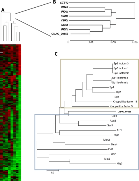
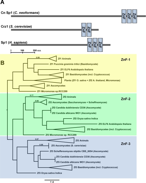
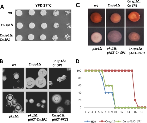
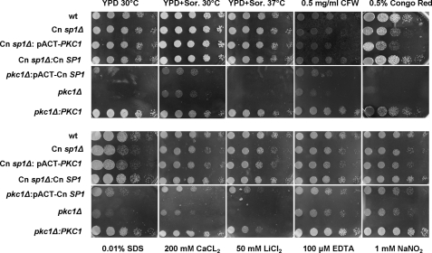
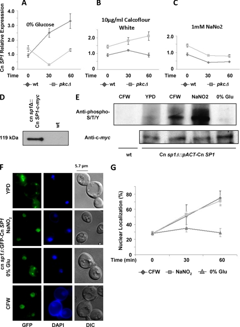
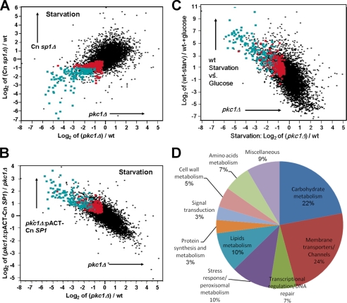
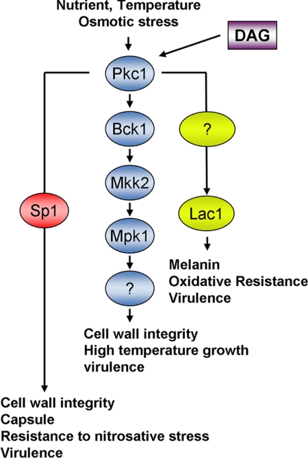
Eukaryotic cells utilize complex signaling systems to detect their environments, responding and adapting as new conditions arise during evolution. The basidiomycete fungus Cryptococcus neoformans is a leading cause of AIDS-related death worldwide and utilizes the calcineurin and protein kinase C-1 (Pkc1) signaling pathways for host adaptation and expression of virulence. In the present studies, a C-terminal zinc finger transcription factor, homologous both to the calcineurin-responsive zinc fingers (Crz1) of ascomycetes and to the Pkc1-dependent specificity protein-1 (Sp1) transcription factors of metazoans, was identified and named SP1 because of its greater similarity to the metazoan factors. Structurally, the Cryptococcus neoformans Sp1 (Cn Sp1) protein was found to have acquired an additional zinc finger motif from that of Crz1 and showed Pkc1-dependent phosphorylation, nuclear localization, and whole genome epistatic associations under starvation conditions. Transcriptional targets of Cn Sp1 shared functional similarities with Crz1 factors, such as cell wall synthesis, but gained the regulation of processes involved in carbohydrate metabolism, including trehalose metabolism, and lost others, such as the induction of autophagy. In addition, overexpression of Cn Sp1 in a pkc1Δ mutant showed restoration of altered phenotypes involved in virulence, including cell wall stability, nitrosative stress, and extracellular capsule production. Cn Sp1 was also found to be important for virulence of the fungus using a mouse model. In summary, these data suggest an evolutionary shift in C-terminal zinc finger proteins during fungal evolution, transforming them from calcineurin-dependent to PKC1-dependent transcription factors, helping to shape the role of fungal pathogenesis of C. neoformans.


Figures

FIGURE 1.
FIGURE 1.
Transcriptional and protein sequence comparison of a C-terminal zinc finger (CNAG_00156). A, heat map display of a comparative microarray experiment of C. neoformans strains Cn sp1Δ (cnag_00156Δ), pkc1Δ, pka1Δ, cna1Δ, cbk1Δ, ssa1Δ, ste12Δ, and vad1Δ. Strains were grown overnight to mid-log phase and induced in 0% glucose medium for 1 h before RNA extraction. WT (H99) induced under the same conditions was used as reference for each mutant. A cluster analysis of genes significantly altered in mutant strains with p < 0.0001 is presented. B, close-up view of the cluster analysis presented in A. C, neighbor-joining phylogenic analysis of the Cn Sp1 (Cnag_00156) with S. cerevisiae (blue frame) and H. sapiens (tan frame) C-terminal zinc finger transcription factors.
FIGURE 2.
FIGURE 2.
C. neoformans and the H. sapiens Sp1 share similar functional domains. A, the SMART-identified zinc finger domain pattern, showing Cn Sp1, Crz1, and H. sapiens Sp1 zinc finger domains (ZnF). B, phylogenetic analysis of metazoan and fungal zinc finger motifs (ZnF-1 to -3) of C. neoformans. Parsimonious trees were constructed using the heuristic parsimony algorithm of PAUP 4b10, as described under “Experimental Procedures.” Clades consisting of major taxonomic groups were collapsed when possible. Bootstrap support percentages greater than 70% are shown above the branches.
FIGURE 3.
FIGURE 3.
Cn sp1Δ and pkc1Δ have altered virulence factors. A, WT (H99), Cn sp1Δ, and Cn sp1Δ::Cn SP1 strains were grown on YPD media and suspended in PBS to an A600 of 0.1. Five 5-fold dilutions of each strain were spotted on YPD agar plates, incubated in 37 °C, and observed for 48 h. B, WT, Cn sp1Δ, pkc1Δ, Cn sp1Δ:Cn SP1, pkc1Δ::pACT-Cn SP1, and Cn sp1Δ::pACT-PKC1 strains were grown on YPD plus sorbitol media, stained with India ink, and observed under microscopy. C, laccase activity. The above strains were plated following glucose starvation on asparagine plus neuroepinephrine media and incubated overnight. D, virulence of WT (H99), Cn sp1Δ, and Cn sp1Δ::Cn SP1 strains was evaluated in a mice model following intravenous injection of 106 cfu. *, p < 0.01 for WT versus Cn sp1Δ and Cn sp1Δ::Cn SP1 versus Cn sp1Δ; p > 0.05 for WT versus Cn sp1Δ::Cn SP1.
FIGURE 4.
FIGURE 4.
Cn sp1Δ and pkc1Δ share cell wall integrity defects. WT (H99), Cn sp1Δ, pkc1Δ, Cn sp1Δ::Cn SP1, pkc1Δ::PKC1, pkc1Δ::pACT-Cn SP1, and Cn sp1Δ::pACT-PKC1 strains were grown on YPD plus sorbitol media and suspended in PBS medium to an A600 of 0.1. Five 5-fold dilutions of each strain were spotted on YPD plus 1 m sorbitol agar plates with the various additives as described and incubated at 30 °C (except for the 37 °C experiment) for 48 h. CFW, calcofluor white.
FIGURE 5.
FIGURE 5.
Cn Sp1 is regulated by Pkc1. A–C, Cn SP1 expression was assayed in WT and pkc1Δ strains, before (mid-log phase in YPD medium) and following the detailed conditions by quantitative RT-PCR. Relative expression was calculated using ACT1 as a reference gene. D, c-myc-tagged Cn Sp1 (Cn sp1Δ::pACT-Cn SP1) and WT strains were grown to mid-log phase in YPD and subjected to immunoprecipitation with anti c-myc antibody followed by Western blotting with anti c-myc Ab. E, Cn sp1Δ::pACT-Cn SP1 cells were grown to mid-log phase and immunoprecipitated following a 1-h induction in medium under the indicated conditions (detailed under “Experimental Procedures”). Western blotting was done with either anti-phosphorylated Thr/Ser/Tyr or anti-c-myc as a loading control. Immunoprecipitated WT cells were used as a negative control. F, Cn sp1Δ::GFP-Cn SP1 strains were grown to mid-log phase and induced for 1 h in medium under the indicated conditions (detailed under “Experimental Procedures”). Representative pictures of Cn sp1Δ::GFP-Cn SP1 cells before and following 1-h induction are presented. DAPI was used for nuclear co-staining. G, percentage of nuclear localization of GFP-Cn SP1 before and after 30 and 60 min of induction (n = 300 cells). CFW, calcofluor white; DIC, differential interference contrast.
FIGURE 6.
FIGURE 6.
The Cn Sp1/Pkc1 pathway regulates gene expression following glucose starvation. A, transcriptional profile of Cn sp1Δ (y axis) and pkc1Δ (x axis) compared with WT following glucose starvation. Red dots, genes that are significantly down-regulated in Cn sp1Δ and pkc1Δ (false discovery rate <0.05) compared with WT. Green dots, a subset of the genes labeled in red that are also down-regulated by at least 2-fold compared with WT (n = 163). The genes in these groups are also presented in subsequent panels. B, transcriptional profile of pkc1Δ::pACT-Cn SP1 (compared with pkc1Δ) and pkc1Δ (compared with WT) following glucose starvation, presented on the y and x axis, respectively. Green and red, the same transcripts as in A. C, transcriptional profile of WT (pre- and poststarvation) and pkc1Δ (compared with WT in starvation) presented on the y and x axis, respectively. Green and red, the same transcripts as in A. D, pie diagram detailing the functional categories of 59 genes with known function of the 163 genes identified in A (green dots).
FIGURE 7.
FIGURE 7.
Role of Sp1 in the Pkc1 signaling pathway of C. neoformans. Pkc1 phosphorylates a member of the MAPK signaling pathway, Bck1, with subsequent phosphorylation of Mkk2 and the downstream component Mpk1 to mediate cell wall integrity, growth at high temperature, and virulence. Pkc1 also regulates the transcription factor Sp1 by phosphorylation, leading to activation by nuclear translocation as well as by transcriptional mechanisms, leading to regulation of osmoresistance, cell wall integrity, and virulence in parallel with the MAPK pathway as well as a unique pathway leading to resistance to nitrosative stress, capsule production, and caspofungin resistance. Laccase (Lac1) is regulated by Pkc1 via an unknown pathway. DAG, diacylglycerol. Adapted from Gerik et al. (16).

References

    1. Bichile L. S., Gokhale Y. A., Sridhar V., Gill N. H. (2001) J. Assoc. Physicians India 49, 377–378 - PubMed
    1. Sorrell T. C., Chen S. C. A., Phillips P., Marr K. A. (2011) Cryptococcus: From Human Pathogen to Model Yeast, pp. 595–606, American Society for Microbiology Press, Washington, D.C
    1. French N., Gray K., Watera C., Nakiyingi J., Lugada E., Moore M., Lalloo D., Whitworth J. A., Gilks C. F. (2002) AIDS 16, 1031–1038 - PubMed
    1. Park B. J., Wannemuehler K. A., Marston B. J., Govender N., Pappas P. G., Chiller T. M. (2009) AIDS 23, 525–530 - PubMed
    1. Odom A., Muir S., Lim E., Toffaletti D. L., Perfect J., Heitman J. (1997) EMBO J. 16, 2576–2589 - PMC - PubMed

Publication types

MeSH terms