. 2011 Jun 17;286(24):21706-16.
doi: 10.1074/jbc.M111.232272. Epub 2011 Apr 12.

Stabilization of HIV-1 envelope in the CD4-bound conformation through specific cross-linking of a CD4 mimetic

Affiliations

Stabilization of HIV-1 envelope in the CD4-bound conformation through specific cross-linking of a CD4 mimetic

Grégoire Martin et al. J Biol Chem. .

Erratum in

  • J Biol Chem. 2011 Aug 19;286(33):29442. Ramos, Oscar H P [added]

Abstract

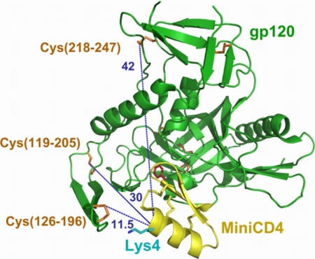
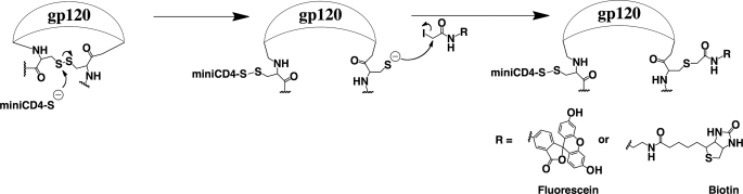
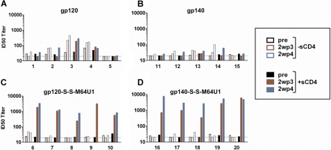
CD4 binding on gp120 leads to the exposure of highly conserved regions recognized by the HIV co-receptor CCR5 and by CD4-induced (CD4i) antibodies. A covalent gp120-CD4 complex was shown to elicit CD4i antibody responses in monkeys, which was correlated with control of the HIV virus infection (DeVico, A., Fouts, T., Lewis, G. K., Gallo, R. C., Godfrey, K., Charurat, M., Harris, I., Galmin, L., and Pal, R. (2007) Proc. Natl. Acad. Sci. U.S.A. 104, 17477-17482). Because the inclusion of CD4 in a vaccine formulation should be avoided, due to potential autoimmune reactions, we engineered small sized CD4 mimetics (miniCD4s) that are poorly immunogenic and do not induce anti-CD4 antibodies. We made covalent complexes between such an engineered miniCD4 and gp120 or gp140, through a site-directed coupling reaction. These complexes were recognized by CD4i antibodies as well as by the HIV co-receptor CCR5. In addition, they elicit CD4i antibody responses in rabbits and therefore represent potential vaccine candidates that mimic an important HIV fusion intermediate, without autoimmune hazard.


Figures

FIGURE 1.
FIGURE 1.
Distance determination between accessible disulfide bonds on gp120 and the α-carbon on position 4 of the miniCD4. This picture was adapted from Stricher et al. (55), using PyMOL. MiniCD4 and gp120 are depicted in yellow and green, respectively. Disulfide bonds are shown in sticks (yellow for miniCD4 and orange for gp120). The three solvent-accessible cystines on gp120 are numbered. His4 was changed to lysine (cyan). The critical Phe23 residue of the miniCD4 is depicted as red sticks. Blue dotted lines show the distances in angstroms between the α-carbon of the reactive residue on the miniCD4 (Lys4) and the accessible disulfides on gp120.
FIGURE 2.
FIGURE 2.
A, cross-linking analysis by SDS-PAGE. Uncross-linked or cross-linked complexes (lanes 1 and 4, respectively), preincubated in 6 m urea (lanes 2 and 5, respectively) or 8 m urea (lanes 3 and 6, respectively), were analyzed by SDS-PAGE in the presence (lanes 7 and 8) or not (lanes 1–6) of 10% β-mercaptoethanol. M64U1(Biot)-SH cross-linking was revealed by chemiluminescence through its biotin moiety via streptavidin HRP. B, protection experiment showing that M64U1(Biot)-SH is specifically covalently bound onto gp120. Separation on SDS-PAGE of gp120 preincubated or not with 100 eq of sCD4 (lanes 1 and 2, respectively) before mixing with 1 eq of biotinylated M64U1-SH. MiniCD4 binding was revealed through its biotin moiety as described above. Small quantities of labeled gp120 aggregates can be seen at about 250 kDa.
SCHEME 1.
SCHEME 1.
Modification of the released cysteine on gp120. To demonstrate that M64U1-SH cross-linked gp120 through reaction on a disulfide bond and to quantify the cross-linking yield, we coupled 5-(iodoacetamido)fluorescein to the gp120-S-S-M64U1 complex. The released cysteine on gp120 was also coupled to N-biotinoyl-N′-iodoacetamidoethylenediamine for the labeling mapping analysis by mass spectrometry.
FIGURE 3.
FIGURE 3.
HPLC profiles of fluorescently labeled cross-linked complexes. gp120 was reacted with various amounts of M64U1-SH (0 eq, green; 0.5 eq, orange; 1 eq, blue; 3 eq, black) for 30 min. The cross-linked complexes were then incubated with a large excess of 5-iodoacetamidofluorescein, for 45 min, and injected on an analytical Vydac C4 reverse phase column (5 μm, 4.6 × 150 mm; flow rate, 1.5 ml·min−1; solvent A, H2O, 0.1% trifluoroacetic acid; solvent B, acetonitrile; gradient, 30–30% solvent B in 10 min, then 30–50% solvent B in 20 min, and 50–70% solvent B in 5 min). Absorbance was measured at 278 nm (solid lines), and the fluorescence profile was obtained for a 500-nm excitation wavelength and a 522-nm emission wavelength (dotted lines). Note that chromatograms were focused on the area of interest.
FIGURE 4.
FIGURE 4.
Labeled residues mapping by mass spectrometry. A, identification of CAM- and Biot-Cys-modified fragment 122–135-residue sequence by PSD MS/MS analysis of precursor ion at m/z 1939.00. Daughter ions were assigned by b- and y-ions series. Two sequences were identified as follows: LTPLCBiotVTLHCCAMTNLK (top) and LTPLCCAMVTLHCBiotTNLK (bottom). B, identification of CAM- and Biot-Cys-modified fragment 193–207-residue sequence by PSD MS/MS analysis of precursor ion at m/z 1988.96. Daughter ions were assigned by b- and y-ions series. The unique sequence identified was LINCBiotDTSVITQACCAMPK.
FIGURE 5.
FIGURE 5.
Binding analysis of the Env-S-S-M64U1 complexes to the 48d CD4i mAb. M64U1-SH or sCD4 (control) was incubated with gp120 or gp140, and CD4-induced epitope induction was evaluated by surface plasmon resonance. Experiments were conducted in HBS (HEPES-buffer saline, 3 mm EDTA, 0.05% Biacore surfactant, pH 7.4). Each sample was analyzed at 10 nm. Sample binding was tested on immobilized CD4i 48d mAb. Dashed lines, gp120 or gp140; dotted lines, M64U1-SH + (gp120 or gp140); solid lines, sCD4 + (gp120 or gp140). A, gp120 data; B, gp140 data.
FIGURE 6.
FIGURE 6.
Immunochemical characterization of the gp140-S-S-M64U1 complex compared with gp140. Serial dilutions of gp140 (filled square) and gp140-S-S-M64U1 (open square) were preincubated on immobilized lectin (concanavalin A) and incubated with several mAbs as follows: 2G12 (A), 2F5 (B), 447–52D (C), MN215 (D), X5 (E), 17b (F), 48d (G), and b12 (H). Anti-gp120 mAbs binding was finally detected with peroxidase-labeled secondary antibodies.
FIGURE 7.
FIGURE 7.
gp120-S-S-M64U1 binding to CCR5 chemokine receptor determined by flow cytometry. gp120 (2nd lane) and gp120 preincubated with either sCD4 (3rd and 7th lanes), M48U1 (4th and 8th lanes), or M64U1-SH (5th and 9th lanes) were incubated at 3 nm on cells overexpressing the CCR5 receptor. Each complex was also tested for cell surface binding after treatment with 3 eq of X5 CD4i antibody (6th to 9th lanes). gp120 binding to CCR5 was detected by using D7324 anti-gp120 mAb and a secondary antibody labeled with a phycoerythrin moiety. Background was evaluated by binding of D7324 on CCR5 in absence of gp120 (1st lane). Data are shown in full bars for gp120 full-length and in open bars for gp120ΔV2 (as a substitute of gp140ΔV2).
FIGURE 8.
FIGURE 8.
CD4i antibody titer. Rabbits were immunized with gp120, gp140, or with M64U1-SH cross-linked to these two envelopes. Four protein immunizations were administered intramuscularly, in the gluteus, at weeks 0, 4, 12, and 24. Serum samples were collected before the first immunization (pre) and 2 weeks post third (2wp3) and fourth (2wp4) immunizations and titered for their CD4i antibodies. The 50% inhibition dose (ID50) for each serum corresponds to the CD4i antibody titer that neutralizes 50% of HIV-27312A/V434M in absence (open bars) or in presence (full bars) of sCD4. Note that rabbit number 9 expired 5 days before the fourth boost injection.

References

    1. Cohen J. (2003) Science 302, 1309–1310 - PubMed
    1. Burton D. R., Desrosiers R. C., Doms R. W., Koff W. C., Kwong P. D., Moore J. P., Nabel G. J., Sodroski J., Wilson I. A., Wyatt R. T. (2004) Nat. Immunol. 5, 233–236 - PubMed
    1. Gallo R. C. (2005) Lancet 366, 1894–1898 - PubMed
    1. Girard M. P., Osmanov S. K., Kieny M. P. (2006) Vaccine 24, 4062–4081 - PubMed
    1. Earl P. L., Sugiura W., Montefiori D. C., Broder C. C., Lee S. A., Wild C., Lifson J., Moss B. (2001) J. Virol. 75, 645–653 - PMC - PubMed

Publication types