. 2011 Jun 3;286(22):19724-34.
doi: 10.1074/jbc.M110.205781. Epub 2011 Apr 12.

Scapinin-induced inhibition of axon elongation is attenuated by phosphorylation and translocation to the cytoplasm

Affiliations

Scapinin-induced inhibition of axon elongation is attenuated by phosphorylation and translocation to the cytoplasm

Hovik Farghaian et al. J Biol Chem. .

Abstract

Scapinin is an actin- and PP1-binding protein that is exclusively expressed in the brain; however, its function in neurons has not been investigated. Here we show that expression of scapinin in primary rat cortical neurons inhibits axon elongation without affecting axon branching, dendritic outgrowth, or polarity. This inhibitory effect was dependent on its ability to bind actin because a mutant form that does not bind actin had no effect on axon elongation. Immunofluorescence analysis showed that scapinin is predominantly located in the distal axon shaft, cell body, and nucleus of neurons and displays a reciprocal staining pattern to phalloidin, consistent with previous reports that it binds actin monomers to inhibit polymerization. We show that scapinin is phosphorylated at a highly conserved site in the central region of the protein (Ser-277) by Cdk5 in vitro. Expression of a scapinin phospho-mimetic mutant (S277D) restored normal axon elongation without affecting actin binding. Instead, phosphorylated scapinin was sequestered in the cytoplasm of neurons and away from the axon. Because its expression is highest in relatively plastic regions of the adult brain (cortex, hippocampus), scapinin is a new regulator of neurite outgrowth and neuroplasticity in the brain.


Figures
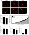
FIGURE 1.
FIGURE 1.
Scapinin inhibits axon elongation in cortical neurons. A, cortical neurons isolated from E18 rats were co-transfected using the Amaxa electroporation system at the time of plating with GFP and scapinin (n = 111) or GFP and empty vector (control; n = 117) and incubated for 3 days. Transfected neurons were identified by detection of GFP (green), whereas neuronal morphology was visualized using an antibody that recognizes the neuronal cytoskeleton-associated protein Tau-1 (red). Arrows indicate abnormally large growth cones on scapinin-transfected neurons. Neuronal morphology and neurite lengths were analyzed using the ImageJ software. B–F, results of analyses are presented as graphs, showing a comparison of averaged axon lengths (B), individual axon lengths (C), number of neurites per cell (D), branching from the axon (E), and percentage of neurons with abnormally large growth cones (F). Error bars = S.E., *, p < 0.05, n.s. = not significant, Student's t test.
FIGURE 2.
FIGURE 2.
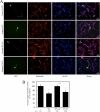
Scapinin is predominantly located in the axon and nucleus. A, immunofluorescence microscopy of endogenous scapinin in 3 DIV rat cortical neurons using two different polyclonal antibodies to scapinin (green). S-18 recognizes an epitope between amino acids 209 and 259 in both long and short isoforms of scapinin. The N-20 antibody recognizes 20 amino acids at the N terminus of the long form of scapinin that is not present in the short form. Neurons were also stained for phalloidin (red) and DAPI (blue). B, relative intensities of staining for the S-18 and N-20 antibodies between different subcellular locations were measured using the ImageJ software and are presented as percentage of total scapinin staining in graphs (error bars = S.E.).
FIGURE 3.
FIGURE 3.
Inhibition of axon elongation by scapinin is dependent on binding to actin. A, cortical neurons isolated from E18 rats were co-transfected using the calcium phosphate method at 1 DIV with GFP and empty vector (control; n = 131), wild type scapinin (n = 138), S277A mutant (n = 144), or S277D (n = 128) mutant and incubated for 2 days. Transfected neurons were identified by detection of GFP (green), whereas neuronal morphology was visualized using an antibody that recognizes the neuronal somato-dendritic marker protein MAP2 (blue) and phalloidin (red). Neuronal morphology and neurite lengths were analyzed using the ImageJ software. B, average axon lengths are presented as a graph (error bars = S.E., *, p < 0.05, n.s. = not significant, Student's t test).
FIGURE 4.
FIGURE 4.
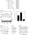
Scapinin is phosphorylated at Ser-277. A, sequence alignment of residues surrounding Ser-277 (bold) of scapinin from various species. Highly conserved proline and basic residues (lysine, arginine) that conform to a Cdk5 phosphorylation consensus sequence are underlined. For comparison, residues of human CRMP2 surrounding Ser-522, which is a confirmed physiological target of Cdk5, are shown. B, scapinin was cloned into a mammalian expression vector, expressed in HEK293 cells, and pulled down via an N-terminal FLAG tag. The isolated protein was separated into two groups; one was left untreated, whereas the other was dephosphorylated using PP1. Following removal of PP1 by washing, both treated and untreated scapinin proteins were subjected to an in vitro kinase assay with recombinant Cdk5/p35 in the presence of radiolabeled ATP. Assays were subjected to SDS-PAGE and visualized by exposure to autoradiography film (32P incorporation; upper panel) or staining with CBR-250 (loading control; lower panel). In addition, the S277A mutant was subjected to the same assay. Arrows indicate the scapinin band. C, the stoichiometry of phosphorylation for untreated, PP1-treated, and S277A mutant forms of scapinin were determined and presented as a graph (error bars = S.E., *, p < 0.05, Student's t test). D, wild type and mutant forms of scapinin were expressed in HEK293 cells, pulled down via their N-terminal FLAG tags, subjected to SDS-PAGE, and transferred to nitrocellulose. Membranes were probed with antibodies that recognize pSer277 (upper panel) or FLAG as a loading control (lower panel). E, lysates from cortex of wild type and Cdk5−/− embryonic mice were subjected to Western blot analysis using antibodies that recognize pSer277 (upper panel) and actin as a loading control (lower panel). F, cultured primary rat cortical neurons were treated with DMSO (control (Con)), 20 μm roscovitine (Rosco), or 2 μm harmine for 48 (upper panel) and 72 h (lower panel). Lysates were prepared and subjected to Western blot analysis using an antibody that recognizes pSer277.
FIGURE 5.
FIGURE 5.
Phosphorylation of Ser-277 on scapinin restores normal axon elongation. A, cortical neurons isolated from E18 rats were co-transfected using the Amaxa electroporation system at the time of plating with GFP and empty vector (control; n = 142), wild type scapinin (n = 158), S277A mutant (n = 140), or S277D mutant (n = 154) and incubated for 3 days. Transfected neurons were identified by detection of GFP (green), whereas neuronal morphology was visualized using an antibody that recognizes the neuronal cytoskeleton-associated protein Tau-1 (red). B, neurons transfected with wild type or S277A scapinin that have abnormally large growth cones are shown by arrows. Neuronal morphology and neurite lengths were analyzed using the ImageJ software. C–F, results of analyses are presented as graphs, showing a comparison of averaged axon lengths (C), average number of neurites per cell (D), branching from the axon (E), and percentage of neurons with abnormally large growth cones (F). Error bars = S.E., *, p < 0.05, n.s. = not significant, Student's t test.
FIGURE 6.
FIGURE 6.
Phosphorylation of scapinin at Ser-277 does not affect actin binding. A, HEK293 cells were transfected with vector only (Control), wild type, S277A, or S277D forms of scapinin, and lysates were prepared. Scapinin was pulled down from the lysates using anti-FLAG-agarose, co-precipitating proteins were subjected to SDS-PAGE, and the gel was stained with CBR-250. The bands at Mr 42,000 were confirmed as actin by Western blot analysis (data not shown). B, the relative intensities of scapinin and actin bands were quantified using densitometry and are presented as a graph (n = 4, error bars = S.E., n.s. = not significant, Student's t test).
FIGURE 7.
FIGURE 7.
Phospho-Ser-277 localizes to the cell body and nucleus. A, immunofluorescence microscopy of endogenous scapinin in 3 DIV rat cortical neurons using polyclonal antibodies that recognize pSer277 (green) or total scapinin (S-18, blue). Neurons were also stained for phalloidin (red) and DAPI (white). B, the ratio of pSer277:total scapinin (S-18) in different subcellular locations is presented as a graph.
FIGURE 8.
FIGURE 8.
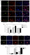
Phospho-mimetic mutant S277D localizes to the cytoplasm and nucleus. A, cortical neurons were transfected using the calcium phosphate method at 1 DIV with GFP only (Control (GFP)), wild type scapinin (n = 21), S277A mutant (n = 21), or S277D mutant (n = 20) and incubated for 2 days. Transfected neurons were identified by detection of GFP (green), whereas neuronal morphology was visualized using an antibody that recognizes the somato-dendritic marker protein MAP2 (blue) and phalloidin (red). Arrows indicate the location of axon. B, relative intensities of wild type and mutant scapinin in different subcellular locations were analyzed using the ImageJ software and are presented as a graph (error bars = S.E., *, p < 0.05, n.s. = not significant, Student's t test). C, immunofluorescence microscopy of endogenous scapinin in 3 DIV and 14 DIV rat cortical neurons using polyclonal antibodies that recognize pSer277 (green) or total scapinin (S-18, blue). Neurons were also stained for phalloidin (red) and DAPI (white). Arrows indicate the location of the nucleus. D, the ratio of pSer277:total scapinin (S-18) in different subcellular locations is presented as a graph.
FIGURE 9.
FIGURE 9.
Actin depolymerization does not change scapinin localization. Rat cortical neurons (3 DIV) were treated with DMSO (Control), 2 μm latrunculin A (LatA), or 1 μm cytochalasin D (CytoD) for 24 h. The subcellular localization of endogenous scapinin was analyzed using immunofluorescence microscopy using polyclonal antibodies that recognize pSer277 (green) or total scapinin (S-18, blue). Neurons were also stained for phalloidin (red) and DAPI (white).

Similar articles

Cited by

References

    1. Sagara J., Higuchi T., Hattori Y., Moriya M., Sarvotham H., Shima H., Shirato H., Kikuchi K., Taniguchi S. (2003) J. Biol. Chem. 278, 45611–45619 - PubMed
    1. Favot L., Gillingwater M., Scott C., Kemp P. R. (2005) FEBS Lett. 579, 100–104 - PubMed
    1. Worch S., Fiedler E., Hansmann I., Schlote D. (2006) Cytogenet. Genome Res. 115, 23–29 - PubMed
    1. Sagara J., Arata T., Taniguchi S. (2009) PLoS One 4, e4247. - PMC - PubMed
    1. Allen P. B., Greenfield A. T., Svenningsson P., Haspeslagh D. C., Greengard P. (2004) Proc. Natl. Acad. Sci. U.S.A. 101, 7187–7192 - PMC - PubMed

Publication types

MeSH terms

LinkOut - more resources