. 2011 Apr 15;71(8):2871-81.
doi: 10.1158/0008-5472.CAN-10-0552. Epub 2011 Apr 12.

In vivo inhibition of human CD19-targeted effector T cells by natural T regulatory cells in a xenotransplant murine model of B cell malignancy

Affiliations

In vivo inhibition of human CD19-targeted effector T cells by natural T regulatory cells in a xenotransplant murine model of B cell malignancy

James C Lee et al. Cancer Res. .

Abstract

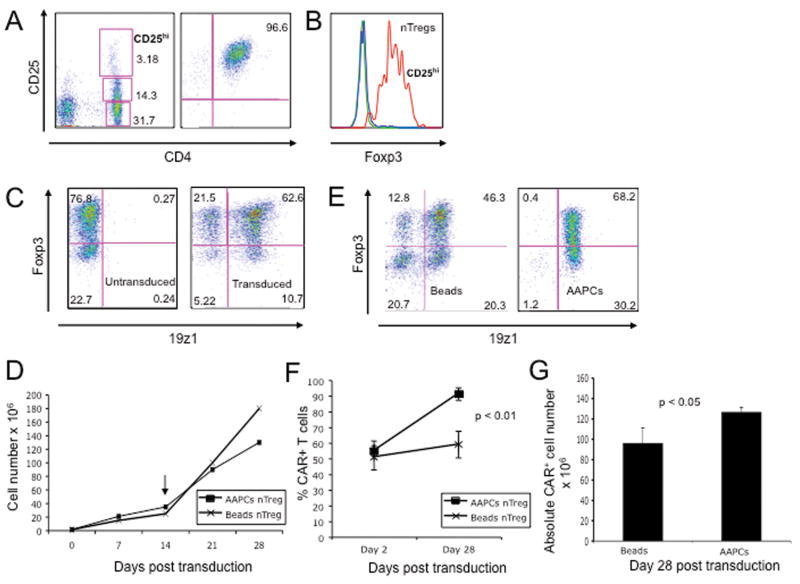
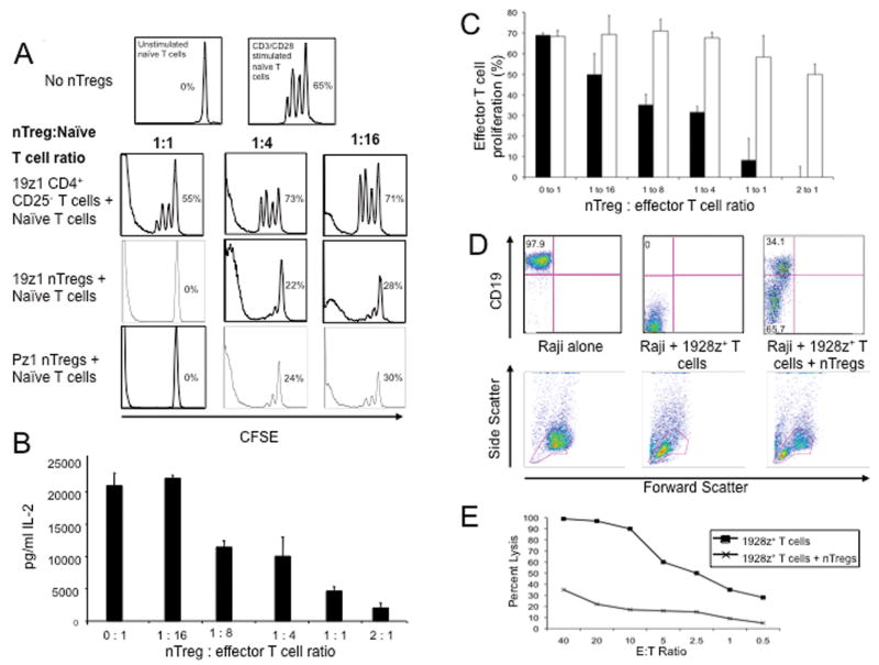
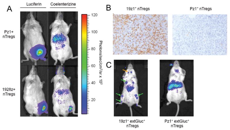
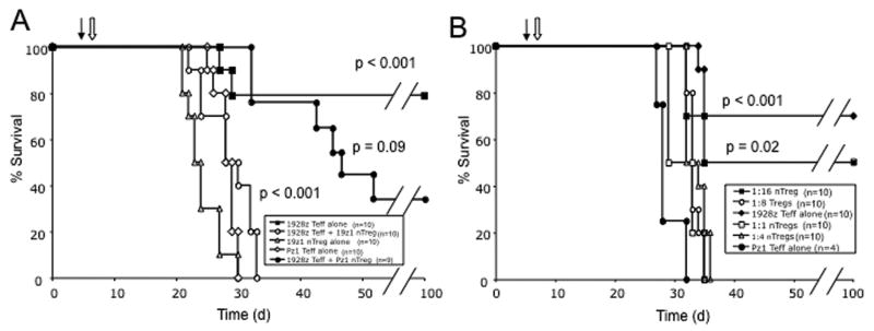
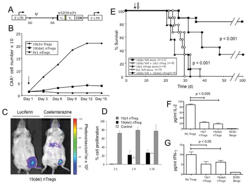
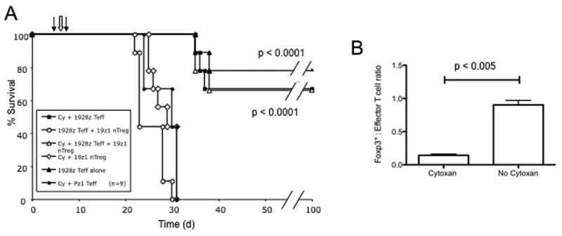
Human T cells genetically modified to express chimeric antigen receptors (CAR) specific to the B cell tumor antigen CD19 can successfully eradicate systemic human CD19(+) tumors in immunocompromised SCID (severe combined immunodeficient)-Beige mice. However, in the clinical setting, CD4(+) CD25(hi) T regulatory cells (Treg) present within the tumor microenvironment may be potent suppressors of tumor-targeted effector T cells. In order to assess the impact of Tregs on CAR-modified T cells in the SCID-Beige xenotransplant model, we isolated, genetically targeted and expanded natural T regulatory cells (nTreg). In vitro nTregs modified to express CD19-targeted CARs efficiently inhibited the proliferation of activated human T cells, as well as the capacity of CD19-targeted 19-28z(+) effector T cells to lyse CD19(+) Raji tumor cells. Intravenous infusion of CD19-targeted nTregs into SCID-Beige mice with systemic Raji tumors traffic to sites of tumor and recapitulate a clinically relevant hostile tumor microenvironment. Antitumor efficacy of subsequently infused 19-28z(+) effector T cells was fully abrogated as assessed by long-term survival of treated mice. Optimal suppression by genetically targeted nTregs was dependent on nTreg to effector T-cell ratios and in vivo nTreg activation. Prior infusion of cyclophosphamide in the setting of this nTreg-mediated hostile microenvironment was able to restore the antitumor activity of subsequently infused 19-28z(+) effector T cells through the eradication of tumor-targeted nTregs. These findings have significant implications for the design of future clinical trials utilizing CAR-based adoptive T-cell therapies of cancer.


Figures

Figure 1
Figure 1. Efficient transduction and expansion of CAR+ nTregs
(A) FACS analysis of nTregs isolated from peripheral blood utilizing the Dynal Regulatory T Cell Isolation Kit (Invitrogen). (B) FACS analyses of isolated nTregs as assessed by intracellular staining for Foxp3 expression. Blue line represents isotype control, green line represents CD4+ CD25- non-Treg control and red line represents nTregs. (C) FACS analyses for Foxp3 and 19z1 expression of isolated nTregs at day 7 following isolation and 19z1 CAR retroviral gene transfer. Similar gene transfer was obtained for nTregs transduced with the 19-28z and Pz1 control CARs (data not shown). (D) nTreg expanded following activation with Dynal CD3/CD28 Human Treg Expander beads (day 0), transduction with the 19z1 CAR, and restimulation (arrow, day 14) with either Expander beads or 3T3(hCD19/CD80) AAPCs. Cell counts, normalized to total cell number, show similar expansion between the AAPC and CD3/CD28 bead activated groups. (E) Foxp3 expression is largely retained following restimulation with either Dynal CD3/CD28 Human Treg Expander beads or 3T3(hCD19/CD80) AAPCs, with the latter population demonstrating enhanced CAR expression. (F) Percentage of expanded T cells expressing the CAR is increased following stimulation on 3T3(hCD19/CD80) AAPCs, but stable following stimulation with Dynal CD3/CD28 Human Treg Expander beads (p < 0.01). (G) Absolute numbers of 19z1+ Tregs is increased following expansion on 3T3(hCD19/CD80) AAPCs when compared to expansion with Dynal CD3/CD28 Human Treg Expander beads.
Figure 2
Figure 2. CAR+ nTregs inhibit expansion of activated naïve T cells, and cytotoxicity of 19-28z+ effector T cells
(A) CSFE-labeled naïve T cells co-cultured with 19z1+ nTregs, Pz1+ nTregs, or 19z1+ Foxp3- CD4+ CD25- T cells at titrated effector to suppressor ratios were activated with CD3/CD28 Human T cell Expander beads. CFSE+ T cells were analyzed via flow cytometry on day 3 post activation. Percent proliferation was calculated using FlowJo software. 19z1+ and Pz1+ nTregs, in contrast to control non-Tregs, efficiently suppressed proliferation of naïve T cells. (B) 19z1+ nTregs inhibit secretion of IL-2 by activated naïve T cells in a dose dependent manner, as assessed by Luminex-based analyses of tissue culture supernatants at 24 hours post co-culture. (C) CFSE-labeled 19-28z+ effector T cells co-cultured with 19z1+ nTregs (solid bars) or Foxp3- CD4+ CD25- T cells at varying effector to suppressor ratios were activated on 3T3(hCD19/CD80) AAPCs for 3 days. 19z1+ nTregs, but not control non-Tregs, suppressed proliferation of 19-28z+ effector T cells. (D) 19z1+ nTregs inhibit 19-28z+ effector T cell cytotoxicity. CD19+ Raji tumor cells were co-cultured at a 1:1:1 ratio with 19-28z+ effector T cells and 19z1+ nTregs. At 24 hours, persistence of Raji tumor cells was assessed by FACS with corresponding FSC/SSC plots provided. Persistence of Raji tumor cells is evident when 19-28z+ effector T cells were co-cultured with 19z1+ nTregs, in contrast to coculture with 19-28z+ effector T cells in the absence of nTregs. (E) Standardized cytotoxicity assay of CD19+ Raji tumor cells co-cultured with 19-28z+ effector T cells alone or 19-28z+ effector T cells with 19z1+ nTregs (at a 1:1 ratio) at various effector to target (E:T) ratios. E:T ratios represent the ratio of 19-28z+ effector T cells to target Raji tumor cells.
Figure 3
Figure 3. CD19-targeted CAR+ nTregs traffic to CD19+ Raji tumor cells in vivo
(A) Dual bioluminescent imaging of Raji tumor and CAR+ nTregs show trafficking of 19z1+ nTregs, but not Pz1+ nTregs, to subcutaneous Raji tumors at 24 hours. SCID-Beige mice were injected subcutaneously with Raji(GFP-FFLuc) cells and 10 days later, with 19z1+ extGLuc+ or control Pz1+ extGLuc+ nTregs. Tumor cells were imaged using the FFLuc specific luciferin substrate, while T cells were imaged using the GLuc specific coelenterazine substrate. (B) Immunohistochemistry staining with an anti-human CD3 antibody confirms the presence of 19z1+ nTregs, but not Pz1+ nTregs, within Raji tumor microenvironment. (C) Bioluminescent imaging CAR+ nTregs show trafficking of 19z1+ nTregs, but not Pz1+ nTregs, to systemic Raji tumors at 24 hours, with predicted signal in the bone marrow of the femurs, tibia, and humeri (green arrows) of 19z1+ extGluc+ nTreg infused mice, as well as infiltration of these nTregs into the submandibular lymphnodes (orange arrow).
Figure 4
Figure 4. CD19-targeted nTregs within the Raji tumor microenvironment suppress 19-28z+ effector T cell function in vivo
(A) SCID-Beige mice were injected i.v. with Raji tumor cells on day 0, followed by CAR+ nTregs on day 5 (filled arrow) and CAR+ effector T cells on day 6 (open arrow). 19z1+ nTregs fully abrogated eradication of systemic Raji tumors by 19-28z+ effector T cells as assessed by survival over time when compared to mice treated with 19-28z+ effector T cells alone (p < 0.001). Pz1+ nTregs did not demonstrate significant suppression (p = 0.09 when compared to the 19-28z+ effector T cells alone cohort; p < 0.001 compared to the 19z1+ nTreg plus 19-28z+ effector T cells cohort). Data represents combined results from 2 independent experiments. (B) 19z1+ nTregs inhibited effector 19-28z+ T cells in a dose dependent manner with infused nTreg to effector T cell ratios of 1:1, 1:4, and 1:8 resulting in no long term surviving mice (all with p < 0.001, compared to 19-28z Teff alone cohort) while a 1:16 nTreg to effector T cell ratio allowed for a 50% long-term survival of treated mice (p = 0.02, compared to Pz1+ Teff treated control cohort). Survival of the 1:16 nTreg to effector T cell treated cohort was statistically similar to the 19-28z Teff alone control cohort (p = 0.3). Similar results were obtained in tumor bearing mice following prior infusion with 19-28z+ nTregs (data not shown). d: days since Raji tumor cell injection.
Figure 5
Figure 5. Optimal in vivo nTreg suppression is dependent on activation of nTregs within the tumor microenvironment
(A) Schematic of the 19(del) CAR. Black box, CD8 leader sequence; green box, (Gly3Ser)4 linker; LTR, long terminal repeat; SD, splice donor; SA, splice acceptor; arrows, start of transcription. (B) 19(del)+ nTregs, like control Pz1+ nTregs, but in contrast to 19z1+ nTregs, fail to expand following co-culture on 3T3(hCD19/CD80) AAPCs consistent with a lack of T cell activation mediated through the 19(del) CAR. (C) 19(del)+ extGLuc + nTregs retain the ability to traffic to Raji tumor in vivo. SCID-Beige mice bearing palpable Raji(GFP-FFLuc) tumors were injected i.v. with 19(del)+ extGLuc+ T cells. Mice were imaged at 24 hours post nTreg infusion. (D) 19(del) + nTregs retain the ability to inhibit expansion of activated naïve T cells. CFSE-labeled naïve T cells co-cultured with 19(del) + Tregs, 19z1+ Tregs, or 19z1+ CD4+ CD25- non-Treg control T cells at varying effector to suppressor ratios were activated with Dynabeads CD3/CD28 Human T cell Expander beads. Both 19(del)+ nTregs and 19z1+ nTregs suppressed proliferation of naïve T cells when compared to the non-Treg control co-cultures. (E) Infusion of Raji tumor bearing mice with 19(del)+ nTregs partially inhibited antitumor efficacy of 19-28z+ effector T cells as assessed by long-term survival. Survival of mice previously infused with 19(del)+ nTregs, when compared to mice treated with 19-28z+ effector T cells alone, was significantly lower (p < 0.001) but significantly improved when compared to mice previously treated with 19z1+ nTregs (p < 0.001). Data represents combined results from 2 independent experiments. Closed arrow: nTreg infusion; open arrow, effector 19-28z+ T cell infusion; d, days since Raji tumor cell injection. At 24 hours following 19-28z+ effector T cell infusion, prior infusion with either 19z1+ or 19(del)+ nTregs significantly reduced serum levels of IL-2 (F) (p < 0.005), and IFNγ (G) (p < 0.05), when compared to mice treated with 19-28z+ effector T cells alone. Figure 5F and 5G represent the combined data from 2 independent experiments with cohorts of 3 mice in each experiment.
Figure 6
Figure 6. Cyclophosphamide lymphodepletion following 19z1+ nTreg infusion, enhances 19-28z+ effector T cell tumor cell eradication, altering the nTreg to effector T cell ratio within the tumor microenvironment
(A) SCID-Beige were mice injected i.v. with Raji tumor cells on day 0, followed by 19z1+ nTregs on day 5 (left filled arrow), i.p. injection of cyclophosphamide (100mg/kg) on day 6 (open arrow), and 19-28z or Pz1+ effector T cells on day 7 (right black arrow). Long term survival comparable to no-nTreg controls was observed in mice injected with 19z1+ nTregs followed by cyclophosphamide therapy and 19-28z+ effector T cell infusion which was significantly superior to similarly treated mice without prior lymphodepletion (p < 0.0001). Data represents combined results from 2 independent experiments. (B) Foxp3+ nTreg to effector cell ratios were assessed in tumor-involved tissues (bone marrow) by FACS at 24 hours following 19-28z+ effector T cell infusion. Prior cyclophosphamide therapy significantly altered the nTreg to effector T cell ratios in favor of the 19-28z+ effector T cells (0.14 with versus 0.9 without lymphodepletion, p < 0.005). Data represent the average from 2 independent experiments each with cohorts of 3 mice per treatment group.

References

    1. Sadelain M, Riviere I, Brentjens R. Targeting tumours with genetically enhanced T lymphocytes. Nat Rev Cancer. 2003;3:35–45. - PubMed
    1. Brentjens RJ, Latouche JB, Santos E, et al. Eradication of systemic B-cell tumors by genetically targeted human T lymphocytes co-stimulated by CD80 and interleukin-15. Nat Med. 2003;9:279–86. - PubMed
    1. Brentjens RJ, Santos E, Nikhamin Y, et al. Genetically targeted T cells eradicate systemic acute lymphoblastic leukemia xenografts. Clin Cancer Res. 2007;13:5426–35. - PubMed
    1. Cheadle EJ, Gilham DE, Hawkins RE. The combination of cyclophosphamide and human T cells genetically engineered to target CD19 can eradicate established B-cell lymphoma. Br J Haematol. 2008;142:65–8. - PubMed
    1. James SE, Orgun NN, Tedder TF, et al. Antibody-mediated B-cell depletion before adoptive immunotherapy with T cells expressing CD20-specific chimeric T-cell receptors facilitates eradication of leukemia in immunocompetent mice. Blood. 2009;114:5454–63. - PMC - PubMed

Publication types

MeSH terms