Review
. 2011;31(6):521-31.
doi: 10.1159/000324328. Epub 2011 Apr 12.

Aquaporins in cerebrovascular disease: a target for treatment of brain edema?

Affiliations
Review

Aquaporins in cerebrovascular disease: a target for treatment of brain edema?

J Badaut et al. Cerebrovasc Dis. 2011.

Abstract

In cerebrovascular disease, edema formation is frequently observed within the first 7 days and is characterized by molecular and cellular changes in the neurovascular unit. The presence of water channels, aquaporins (AQPs), within the neurovascular unit has led to intensive research in understanding the underlying roles of each of the AQPs under normal conditions and in different diseases. In this review, we summarize some of the recent knowledge on AQPs, focusing on AQP4, the most abundant AQP in the central nervous system. Several experimental models illustrate that AQPs have dual, complex regulatory roles in edema formation and resolution. To date, no specific therapeutic agents have been developed to inhibit water flux through these channels. However, experimental results strongly suggest that this is an important area for future investigation. In fact, early inhibition of water channels may have positive effects in the prevention of edema formation. At later time points during the course of disease, AQP is important for the clearance of water from the brain into blood vessels. Thus, AQPs, and in particular AQP4, have important roles in the resolution of edema after brain injury. The function of these water channel proteins makes them an excellent therapeutic target.


Figures

Fig. 1
Fig. 1
Schematic of the AQP homotetramer assembly within the lipid membrane from a lateral (a) and a top view (b) resulting in a central pore permeable to cations and gases (thick arrows). Each individual AQP facilitates bidirectional water movement depending on the osmotic gradient (blue arrows, thin grey in the printed version). mb = Membrane.
Fig. 2
Fig. 2
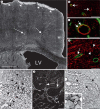
AQP4 distribution within the rodent brain. a AQP4 is observed along intraparenchymal blood vessels (arrows) in the gray matter, ventricles, in the glia limitans (arrowheads) and ependymal cells along the lateral ventricle (LV). b AQP4-immunolabelling (red, arrows) is observed adjacent to the blood vessels but not on endothelial cells, stained by endothelial brain antigen (green). c AQP4-immunolabelling (red, arrows) on the astrocytic endfeet is revealed by anti-glial fibrillary acidic protein staining (green, arrows) within the parietal cortex. d In the corpus callosum, AQP4 expression (red) on astrocyte endfeet adjacent to blood vessels (endothelial brain antigen, green, arrows), cell bodies and processes (arrowheads). e AQP4 distribution in the cerebellum differs from the cortex. AQP4 is observed around the blood vessels (arrowheads) and also around the Purkinje cell bodies (P, *) and neurons of the granular layer (G, arrows). f Higher magnification shows AQP4 expression at the basal membrane (arrows) of the cell bodies of Purkinje neurons (P) in the cerebellum. g, h A similar distribution was observed in the magnocellular hypothalamic nuclei such as in the paraventricular nucleus (dotted line). AQP4 is present on the astrocyte processes in contact with blood vessels (* in h) and the magnocellular neurons in hypothalamic paraventricular nucleus (arrows). Bar: a = 500 μm; b, d–f = 50 μm; c, h = 10 μm.
Fig. 3
Fig. 3
a Association between AQP4-m1 and AQP4-m23 isoforms contributes to form OAPs. Higher expression of AQP4-m23 contributes to the formation of large OAPs. Recent knowledge on AQP led us to hypothesize that the large OAPs contribute to gas and cation diffusion in the astrocyte membranes through central pores (thick arrows). b Increased AQP4-m1, for example after stroke, induces disruption of OAPs (see [32]).
Fig. 4
Fig. 4
AQP4s in cerebrovascular disease and injury. a1, a2 AQP4 expression is increased in the striatum within the future lesion site 30 min after reperfusion in a model of MCA occlusion [22, 32, 35]. a1 Contralateral striatum. a2 Ipsilateral striatum.b AQP4 Western blots confirmed the increase in AQP4 at 30 min after reperfusion in the ispilateral striatum [22, 32, 35]. c AQP4 expression of isosforms (n = 7). Quantification showed an increase in the AQP4-m1 in the ipsilateral (dark grey column) striatum that validates the disruption of the OAPs observed after stroke [22, 32, 35]. Light grey column= Contralateral striatum. d1, d2 AQP4 expression is decreased where T2WI images (d1) showed hyperintense signals suggesting high water content (*) at day 28 after stroke. However, at the border of the lesion (dotted line) high expression of AQP4 correlated with differences in T2 signals (arrows). e Glial fibrillary acidic protein staining (green) revealed a glia scar adjacent to the hyperintense signal from the T2 images (d1). The glia scar is characterized by a high level of AQP4 immunoreactivity (red, arrows). f1, f2 Similar observations were made in the spinal cord after injury in rats (SCI), where AQP4 expression was highly induced 2 months after a lesion. High expression of AQP4 observed around the cyst (yellow arrow) that was confirmed on T2WI (f2) [38]. Bar: a1, e = 50 μm; d1 = 1 mm; f1 = 200 μm.
Fig. 5
Fig. 5
Schematic drawing of the time course of edema formation after transient MCA occlusion (a) highlighting 3 different edema phases: anoxic (b), ionic (c) and vasogenic edema (d). a The first few minutes after an occlusion are characterized by anoxic edema (b) with loss of ionic gradients. The shear stress induced by reperfusion is followed by two peaks of brain swelling observed after transient MCA occlusion, one around 30 min coinciding with ionic edema (c) and a second at 48 h reflecting vasogenic edema (d). The other line indicates changes in the level of AQP4 expression following brain swelling (edema). b The initial anoxic edema is characterized as a swelling of the astrocytes and the neuronal dendrites caused by a disruption of the cellular ionic gradients and the entry of ions followed by water entry leading to cellular swelling through AQP4, transporters and ionic channels. c In ionic edema, astrocytes are still swollen and neuronal death starts, resulting in shrinkage of the neurons and shear stress on the nonperfused vascular tree that results in early transient leakage of the BBB. d Vasogenic edema is a result of disruption of the tight junctions between endothelial cells, leading to increased permeability of the cerebral blood vessels to albumin and other plasma proteins, further contributing to swelling of astrocytes and subsequent neuronal cell death.

References

    1. Klatzo I. Brain oedema following brain ischaemia and the influence of therapy. Br J Anaesth. 1985;57:18–22. - PubMed
    1. Gonen T, Walz T. The structure of aquaporins. Q Rev Biophys. 2006;39:361–396. - PubMed
    1. Yu J, Yool AJ, Schulten K, Tajkhorshid E. Mechanism of gating and ion conductivity of a possible tetrameric pore in aquaporin-1. Structure. 2006;14:1411–1423. - PubMed
    1. Musa-Aziz R, Chen LM, Pelletier MF, Boron WF. Relative CO2/NH3 selectivities of AQP1, AQP4, AQP5, AmtB, and RhAG. Proc Natl Acad Sci USA. 2009;106:5406–5411. - PMC - PubMed
    1. Nielsen S, Nagelhus EA, Amiry-Moghaddam M, Bourque C, Agre P, Ottersen OP. Specialized membrane domains for water transport in glial cells: high-resolution immunogold cytochemistry of aquaporin-4 in rat brain. J Neurosci. 1997;17:171–180. - PMC - PubMed

Publication types

MeSH terms