Left atrial myxoma with papillary fibroelastoma-like features
- PMID: 21487526
- PMCID: PMC3071663
Left atrial myxoma with papillary fibroelastoma-like features
Abstract
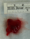
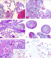
Although rare, papillary fibroelastoma (PFE) of the heart valves and atrial myxoma represent the two most common cardiac tumors. Coexistence of these two lesions has been documented in rare case reports. We describe the case of a 77-year-old man who presented with slightly progressive chest pain associated with dyspnea, fatigue and edema of the lungs. Transthoracic echocardiography detected a left atrial mass that has been successfully excised. Histopathological examination showed a neoplasm combining features of both atrial myxoma and PFE. However, close evaluation of the latter showed microscopic foci of myxomatous tissue within papillary cores, indicating that the PFE-like component has developed around preexisting myxomatous tissue that served as a nidus for papillary fronds, probably by a process of fibrinous microthrombosis, organization and endothelialisation. This unusual case may shed light on the pathogenesis of the PFE pattern.
Keywords: Papillary fibroelastoma; atrium; calretinin; myxoma.
Figures


References
-
- Burke AP, Virmani R. Atlas of Tumor Pathology, third series, fascicle 16. Washington, DC: Armed Forces Institute of Pathology; 1996. Tumors of the heart and great vessels.
-
- Blondeau P. Primary cardiac tumors. French studies of 533 cases. Thorac cardiovasc Surg. 1990;38:192–195. - PubMed
-
- Raeburn C. Papillary fibro-elastic hamartomas of the heart valves. J Pathol Bact. 1953;65:371–373. - PubMed
-
- Strecker T, Agaimy A, Marwan M, Zielezinski T. Papillary fibroelastoma of the aortic valve: appearance in echocardiography, computed tomography, and histopathology. J Heart Valve Dis. 2010;19:812. - PubMed
-
- Prifti E, Bonacchi M, Salica A. Mitral valve myxoma concomitant with papillary fibroelastoma. Ann Thorac Surg. 2000;70:335–6. - PubMed
Publication types
MeSH terms
LinkOut - more resources
Full Text Sources
