Review
. 2011 Mar;4(3):315-21.
Epub 2011 Mar 22.

Metastatic balloon cell malignant melanoma: a case report and literature review

Affiliations
Review

Metastatic balloon cell malignant melanoma: a case report and literature review

Lili Lee et al. Int J Clin Exp Pathol. 2011 Mar.

Abstract

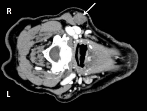
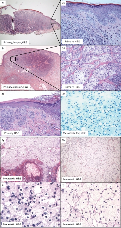
A case of metastatic balloon cell malignant melanoma (BCMM) is presented. The balloon melanoma cells (BMC) were absent in the shave biopsy of the primary lesion and present as a minor component in the wide and deep excision. A subsequent right neck lymph node metastasis showed complete replacement of the lymph node by large, foamy cells. Though the tumor was amelanocytic and Fontana-Masson stain failed to reveal melanin, it stained positively for S-100, HMB-45, and Melan-A. Ultrastructurally, the foamy cells were characterized by cytoplasmic vacuolization and a lack of melanosomes. The differential diagnosis of metastatic balloon cell malignant melanoma is broad, and clinicopathologic correlation may play a critical role in achieving the correct diagnosis.

Keywords: Malignant; balloon cell; clear cell tumors; melanoma.


Figures

Figure 1
Figure 1
CT with contrast of the right neck mass showing a well circumscribed 2.3 × 1.7 × 1.6 cm mass (arrow).
Figure 2
Figure 2
(a)Primary melanoma, shave biopsy: confluent irregular nests within the dermis and as single units at the dermo-epidermal junction (H&E, ×4); (b) Close-up of Figure 2a. No clear cells or melanin pigment was identified (H&E, ×40); (c). Primary melanoma, wide and deep excision: note the minimal amount of balloon cells in a background of predominantly “basaloid” and spindle-shaped melanoma cells. The balloon cells occupy less than 50% of the tumor (H&E, 4×); (d) Primary melanoma, wide and deep excision: the polyhedral balloon cells are arranged in nests and cords separated by thin fibrous septae. They show discernabie cytopiasmic membranes and foamy to clear cytoplasm with occasional pale eosinophilic granules. They are minimally pleomorphic, variably sized, ranging from 40-100 microns in diameter, and show a central or eccentric nuclei. Few prominent nucleoi are seen (H&E, 40×); (e) Primary melanoma, wide and deep excision: intraepidermal pagetoid spread is present. Note the presence of a mitotic figure in the spindle-shaped population of melanoma cells. Hemosiderin is observed (H&E, 40×); (f) Metastatic balloon cell malignant melanoma (BCMM), fine-needle aspiration: sheets of discohesive, large, polyhedral foamy cells with granular cytoplasm. Cytopiasmic membranes are indistinct. These cells show bland nuclear features with a low nuclear to cytopiasmic ratio. Eccentrically placed nuclei are round to ovoid. Occasional intranuclear cytopiasmic inclusions and inconspicuous nucleoli are identified. No pigmentation is present (Pap, 40×); (g) Metastatic BCMM. The lymph node shows near complete replacement by balloon melanoma cells. No spindle-shaped or epithelioid melanoma cells are present H&E, 4×); (h) Metastatic BCMM. The balloon melanoma cells are arranged in nests separated by thin fibrous septae and are histologically identical to those seen in the primary lesion (H&E, 10×); (i) Metastatic BCMM. The tumor shows mild pleomorphism with focal areas of high cellular-ity and nuclear pleomorphism (H&E, 60×); (j) Metastatic BCMM. The majority of the tumor is cytologically bland with minimal pleomorphism (H&E, 60×).
Figure 3
Figure 3
Metastatic balloon cell malignant melanoma (BCMM). (a) Metastatic BCMM, positive for Melan-A (Melan-A, 40×). Inset shows primary melanoma, wide and deep excision: Melan-A showing a positive staining pattern similar to S-100. Transitional cells show darker cytopiasmic staining than balloon melanoma cells (Melan-A, 40×); (b) Metastatic BCMM, positive for S-100. Transitional cells show darker cytopiasmic staining than balloon melanoma cells (S-100, 40×); (c) Metastatic BCMM, positive for HMB-45 (HMB-45, 40×); (d) Metastatic BCMM, negative for CD68, ruling out granular cell tumor and xanthomatous lesion. However, some BCMM may be positive for CD68 (CD68, 40×).

References

    1. Kao GF, Helwig EB, Graham JH. Balloon cell malignant melanoma of the skin. A clinicopathologic study of 34 cases with histochemical, immunohistochemical, and ultrastructural observations. Cancer. 1992;69:2942–2952. - PubMed
    1. Aloi FG, Coverlizza S, Pippione M. Balloon cell melanoma: a report of two cases. J Cutan Pathol. 1988;15:230–233. - PubMed
    1. Gardner WA, Vazquez MD. Balloon Cell Melanoma. Archives of Pathology. 1970;89:470. &. - PubMed
    1. Ranchod M, Path MM. Metastatic Melanoma with Balloon Cell Changes. Cancer. 1972;30:1006. &. - PubMed
    1. Akslen LA, Myking AO. Balloon Cell Melanoma Mimicking Clear Cell-Carcinoma. Pathology Research and Practice. 1989;184:548–550. - PubMed

LinkOut - more resources