

doi: 10.1002/0471140864.ps1210s64.
Preparation and analysis of glycan microarrays
Affiliations
- PMID: 21488041
- PMCID: PMC3097418
- DOI: 10.1002/0471140864.ps1210s64
Item in Clipboard
Preparation and analysis of glycan microarrays
Curr Protoc Protein Sci.
2011 Apr.
Abstract
Determination of the binding specificity of glycan-binding proteins (GBPs), such as lectins, antibodies, and receptors, has traditionally been difficult and laborious. The advent of glycan microarrays has revolutionized the field of glycobiology by allowing simultaneous screening of a GBP for interactions with a large set of glycans in a single format. This unit describes the theory and method for production of two types of glycan microarrays (chemo/enzymatically synthesized and naturally derived), and their application to functional glycomics to explore glycan recognition by GBPs. These procedures are amenable to various types of arrays and a wide range of GBP samples.
© 2011 by John Wiley & Sons, Inc.
Figures
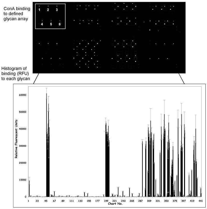
Con A binding to the CFG glycan array. The top panel is an image of fluorescently detected Con A binding to the glycan array slide. The replicates of one set of 6 spots, corresponding to one glycan, are noted. The bottom panel is a histogram of quantified Con A binding to the glycan array, where the x-axis is the chart number (glycan number) and the y-axis is relative fluorescent units (RFU).
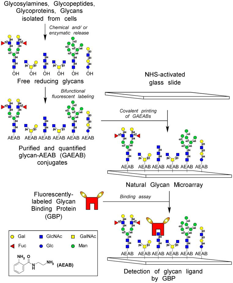
Preparation of natural glycan microarrays. After isolating the glycans from a natural source, they are labeled with a bifunctional fluorescent dye (ex. AEAB), captured on a glass slides, and interrogated by glycan binding proteins (GBPs).
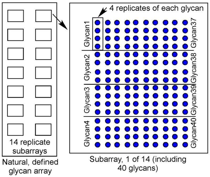
Schematic of natural, defined glycan array printed with 14 identical subarrays per slide. The multi-chamber adaptor can be place over the slide surface to create 14 individual chambers for 14 different, simultaneous assays. In this example, each of the 40 glycans is printed in replicates of 4. In some cases, the printed compounds include controls such as glycopeptides, biotin, etc.

The binding of biotinylated SNA to a natural glycan array containing 52 GAEABs and 4 controls. After obtaining a fluorescent image of the biotinylated SNA bound by cyanine5-streptavidin, binding is quantified and displayed as a histogram of the average RFU bound to each glycan. The 8 glycan structures that were bound by SNA are displayed next to their respective peak.
References
-
- Bohorov O, Andersson-Sand H, et al. Arraying glycomics: a novel bi-functional spacer for one-step microscale derivatization of free reducing glycans. Glycobiology. 2006;16:21C–27C. - PubMed
Publication types
MeSH terms
Substances
Grants and funding
LinkOut - more resources
Full Text Sources
Other Literature Sources
Research Materials
