. 2011 Jul;54(1):273-84.
doi: 10.1002/hep.24359.

Serum and liver iron differently regulate the bone morphogenetic protein 6 (BMP6)-SMAD signaling pathway in mice

Affiliations

Serum and liver iron differently regulate the bone morphogenetic protein 6 (BMP6)-SMAD signaling pathway in mice

Elena Corradini et al. Hepatology. 2011 Jul.

Abstract

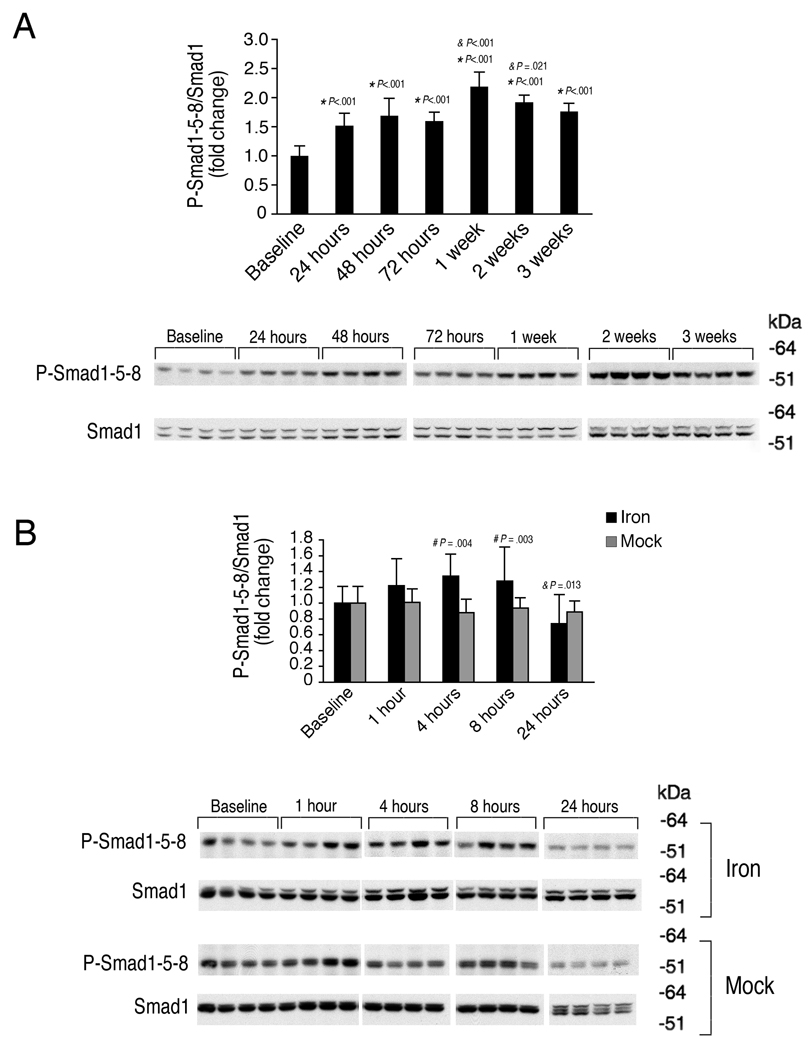
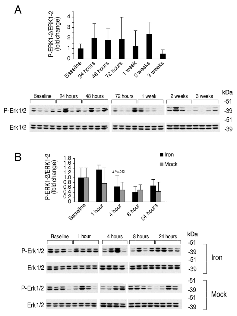
The bone morphogenetic protein 6 (BMP6)-SMAD signaling pathway is a central regulator of hepcidin expression and systemic iron balance. However, the molecular mechanisms by which iron is sensed to regulate BMP6-SMAD signaling and hepcidin expression are unknown. Here we examined the effects of circulating and tissue iron on Bmp6-Smad pathway activation and hepcidin expression in vivo after acute and chronic enteral iron administration in mice. We demonstrated that both transferrin saturation and liver iron content independently influence hepcidin expression. Although liver iron content is independently positively correlated with hepatic Bmp6 messenger RNA (mRNA) expression and overall activation of the Smad1/5/8 signaling pathway, transferrin saturation activates the downstream Smad1/5/8 signaling cascade, but does not induce Bmp6 mRNA expression in the liver. Hepatic inhibitory Smad7 mRNA expression is increased by both acute and chronic iron administration and mirrors overall activation of the Smad1/5/8 signaling cascade. In contrast to the Smad pathway, the extracellular signal-regulated kinase 1 and 2 (Erk1/2) mitogen-activated protein kinase (Mapk) signaling pathway in the liver is not activated by acute or chronic iron administration in mice.

Conclusion: Our data demonstrate that the hepatic Bmp6-Smad signaling pathway is differentially activated by circulating and tissue iron to induce hepcidin expression, whereas the hepatic Erk1/2 signaling pathway is not activated by iron in vivo.


Figures

Figure 1
Figure 1. Chronic dietary iron administration increases serum iron, serum transferrin saturation, liver iron content, and hepatic hepcidin mRNA in mice
Seven-week-old male C57BL/6 mice were placed on a high iron diet (2% carbonyl iron) and were sacrificed at time zero (Baseline), 24 hours, 48 hours, 72 hours, 1 week, 2 weeks, or 3 weeks after initiation of the high iron diet (N = 6 per group). Animals were analyzed for serum iron (panel A), transferrin saturation (Tf sat, panel B), liver iron content (LIC, panel C), and hepcidin (Hamp, also known as Hamp1) relative to Rpl19 mRNA expression by quantitative real-time RT-PCR (panel D). Results are expressed as the mean ± s.d. for serum and tissue iron parameters, and as the mean ± s.d for the fold change compared to the baseline for hepcidin expression. Statistical significance was determined by one-way ANOVA with Holm-Sidak or the Dunnett’s post hoc tests for pair-wise multiple comparisons. A high iron diet significantly increased serum iron (Panel A, F=10.16, P<.001), Tf sat (Panel B, F=65.79, P<.001), LIC (Panel C, F=172.32, P<.001), and Hamp relative to Rpl19 mRNA (Panel D, F=99.40, P<.001). For each group significant changes are shown as (*) for P<.001 in comparison with the baseline, and (&) for P<.001 or as otherwise indicated if significant in comparison with the previous group.
Figure 2
Figure 2. Hamp mRNA levels are independently influenced by both LIC and Tf sat
Seven-week-old male C57BL/6 mice received a standard rodent diet (Baseline), a high iron diet for 1 week (High Fe), or a high iron diet for 1 week followed by a low iron diet (2–6 ppm iron; High Fe then Low Fe) for 24 hours to 8 days as indicated (N = 4 per group). Tissues were analyzed for serum iron (Panel A), Tf sat (Panel B), LIC (Panel C), and Hamp relative to Rpl19 mRNA by quantitative real-time RT-PCR (Panel D). Results are expressed as the mean ± s.d. Statistical significance was determined by one-way ANOVA with Holm-Sidak or the Dunnett’s post hoc tests for pair-wise multiple comparisons. For each group, *P<.001 or as otherwise indicated if significant in comparison to Baseline, #P<.001 or as otherwise indicated if significant in comparison with the High Fe group. A high iron diet significantly increased serum iron, Tf sat, LIC, and Hamp relative to Rpl19 mRNA relative to baseline (Panels A–D). After switching to a low iron diet for 24 hours to 8 days, serum iron and Tf sat were significantly decreased back to baseline levels for all time points, except 8 days, where there was a small but significant reduction in Tf sat from baseline (Panels A–B). After switching to a low iron diet for 24 hours to 8 days, a significantly increased LIC was maintained compared with the baseline group, and the LIC was not significantly decreased from the High Fe group, except for a small but significant decrease at 24 hours (Panel C). After switching to a low iron diet for 24 hours to 8 days, Hamp relative to Rpl19 mRNA levels were decreased to an intermediate level between those achieved by the high iron diet and baseline (Panel D).
Figure 3
Figure 3. Acute enteral iron administration increases serum iron and Tf sat, but not LIC, and induces hepatic Hamp mRNA expression in mice
Nine-week-old male C57BL/6 mice received a single dose of 2 mg elemental iron per kg animal weight (Iron, black bars) or the same volume of water alone by oral gavage (Mock, gray bars). Mice were sacrificed at time 0 (without any treatment, Baseline), and at 1, 4, 8, and 24 hours after either iron or water gavage (N = 6 per group). Animals were analyzed for serum iron (panel A), Tf sat (panel B), LIC (panel C), and Hamp relative to Rpl19 mRNA expression by quantitative real-time RT-PCR (panel D). Results are expressed as the mean ± s.d. for serum and tissue iron parameters, and as the mean ± s.d for the fold change compared to the baseline for hepcidin expression. Statistical significance was determined by two-way ANOVA with Holm-Sidak or the Dunnett’s post hoc tests for pair-wise multiple comparisons. Compared with baseline or mock gavage, a single dose of iron by oral gavage significantly increased serum iron (Panel A, F=3.974, P=0.007 for time, F=25.002, P<.001 for treatment), serum Tf sat (Panel B, F=3.285, P=.018 for time, F=21.223, P<.001 for treatment), and Hamp relative to Rpl19 mRNA (Panel D, F=8.941, P=.001 for time, F=10.678, P=.002 for treatment), but did not affect LIC (Panel C, F=1.60, P=.189 for time, F=.936, P=.338 for treatment). For each iron treated group, significant changes are shown as exact P values for the comparisons with the baseline (*), with the previous group (&), and with the corresponding mock group (#).
Figure 4
Figure 4. Chronic iron administration stimulates hepatic Bmp6 mRNA expression but acute iron administration does not
The animals that underwent chronic iron administration from Figure 1 (Panel A) and acute iron administration from Figure 3 (Panel B) were analyzed for hepatic Bmp6 relative to Rpl19 mRNA expression by quantitative real-time RT-PCR (N = 6 per group). Results are expressed as the mean ± s.d. for the fold change compared to the baseline. Statistical significance was determined by one-way ANOVA (chronic iron administration, Panel A) or two-way ANOVA (acute iron administration, Panel B) with Holm-Sidak or the Dunnett’s post hoc tests for pair-wise multiple comparisons. For each group, significant changes are shown as exact P values for the comparisons with the baseline (*), with the previous group (&), and with the corresponding iron group (#). Hepatic Bmp6 relative to Rpl19 mRNA was significantly increased from baseline by chronic iron administration (Panel A, F=30.20, P<.001), but not by acute iron administration (Panel B), although there was a slight decrease in hepatic Bmp6 relative to Rpl19 mRNA in mock gavage groups compared to the baseline group and the corresponding iron time points (Panel B, F=4.509, P=.003 for time, F=6.99, P=.011 for treatment).
Figure 5
Figure 5. Chronic and acute iron administration both stimulate hepatic Smad1/5/8 signaling
The animals that underwent chronic iron administration from Figure 1 (Panel A) and acute iron administration from Figure 3 (Panel B) were analyzed for hepatic phosphorylated Smad1/5/8 (P-Smad 1-5-8) relative to Smad1 protein by Western blot followed by chemiluminescence quantitation. Results are expressed and statistics analyzed as in Figure 4. Chronic iron treatment significantly increased hepatic P-Smad1/5/8 relative to total Smad1 protein (Panel A, F=21.75, P<.001). Similarly, acute iron administration significantly increased hepatic P-Smad1/5/8 relative to total Smad1 protein compared with the corresponding mock treatment groups, and showed a trend toward a temporal progressive increase compared to baseline (Panel B, F=5.855, P=.02 for treatment, F=2.246, P=.073 for time). For each iron treated group, significant changes are shown as exact P values for the comparisons with the baseline (*), with the previous group (&), and with the corresponding mock group (#).
Figure 6
Figure 6. Chronic and acute iron administration both stimulate expression of hepatic Bmp6-Smad1/5/8 target transcripts Id1 and Smad7
The animals that underwent chronic iron administration from Figure 1 (Panels A–B) and acute iron administration from Figure 3 (Panels C–D) were analyzed for hepatic Id1 (Panels A, C) and Smad7 (Panels B, D) relative to Rpl19 mRNA by quantitative real-time RT-PCR. Results are expressed and statistics analyzed as in Figure 4. Chronic iron treatment significantly increased hepatic Id1 (Panel A, F=27.745, P<.001) and Smad7 relative to Rpl19 mRNA (Panel B, F=7.41, P<.001). Acute iron administration also significantly increased hepatic Id1 relative to Rpl19 mRNA compared with the corresponding mock treatment groups and the baseline (Panel C, F=13.676, P<.001 for treatment, F=4.944, P=.002 for time). Acute iron administration also significantly increased hepatic Smad7 relative to Rpl19 mRNA compared with the corresponding mock groups, although there was only an overall trend toward increased expression compared with the baseline group (Panel D, F=19.208, P<.001 for treatment, F=1.548, P=.203 for time). For each iron treated group, significant changes are shown as exact P values for the comparisons with the baseline (*), with the previous group (&), and with the corresponding mock group (#).
Figure 7
Figure 7. Chronic and acute iron administration have no effect of hepatic Erk1/2 phosphorylation
The animals that underwent chronic iron administration from Figure 1 (Panel A) and acute iron administration from Figure 3 (Panel B) were analyzed for hepatic phosphorylated Erk1/2 (P-ERk1/2) relative to total Erk1/2 protein by Western blot followed by chemiluminescence quantitation. Results are expressed and statistics analyzed as in Figure 4. Chronic iron administration had no significant effect on hepatic P-Erk1/2 relative to total Erk1/2 (Panel A, F=.761, P=.608). Acute iron administration had no significant effect on hepatic P-Erk1/2 relative to total Erk1/2 compared with the corresponding mock gavage groups, although there was a temporal progressive decrease in hepatic P-Erk1/2 relative to Erk1/2 in both the iron and mock gavage groups (Panel B, F=2.246, P=.142 for treatment, F=5.188, P=.002 for time). For each iron treated group, significant changes are shown as exact P values for the comparisons with the baseline (*), with the previous group (&), and with the corresponding mock group (#).

Comment in

Similar articles

Cited by

References

    1. Babitt JL, Lin HY. Molecular mechanisms of hepcidin regulation: implications for the anemia of CKD. Am J Kidney Dis. 2010;55:726–741. - PMC - PubMed
    1. Nemeth E, Tuttle MS, Powelson J, Vaughn MB, Donovan A, Ward DM, et al. Hepcidin regulates cellular iron efflux by binding to ferroportin and inducing its internalization. Science. 2004;306:2090–2093. - PubMed
    1. Pigeon C, Ilyin G, Courselaud B, Leroyer P, Turlin B, Brissot P, et al. A new mouse liver-specific gene, encoding a protein homologous to human antimicrobial peptide hepcidin, is overexpressed during iron overload. J Biol Chem. 2001;276:7811–7819. - PubMed
    1. Nicolas G, Chauvet C, Viatte L, Danan JL, Bigard X, Devaux I, et al. The gene encoding the iron regulatory peptide hepcidin is regulated by anemia, hypoxia, and inflammation. J Clin Invest. 2002;110:1037–1044. - PMC - PubMed
    1. Nemeth E, Valore EV, Territo M, Schiller G, Lichtenstein A, Ganz T. Hepcidin, a putative mediator of anemia of inflammation, is a type II acute-phase protein. Blood. 2003;101:2461–2463. - PubMed

Publication types

MeSH terms