. 2011 Jul;89(7):1001-17.
doi: 10.1002/jnr.22624. Epub 2011 Apr 12.

Spatial and temporal changes in promoter activity of the astrocyte glutamate transporter GLT1 following traumatic spinal cord injury

Affiliations

Spatial and temporal changes in promoter activity of the astrocyte glutamate transporter GLT1 following traumatic spinal cord injury

Angelo C Lepore et al. J Neurosci Res. 2011 Jul.

Abstract

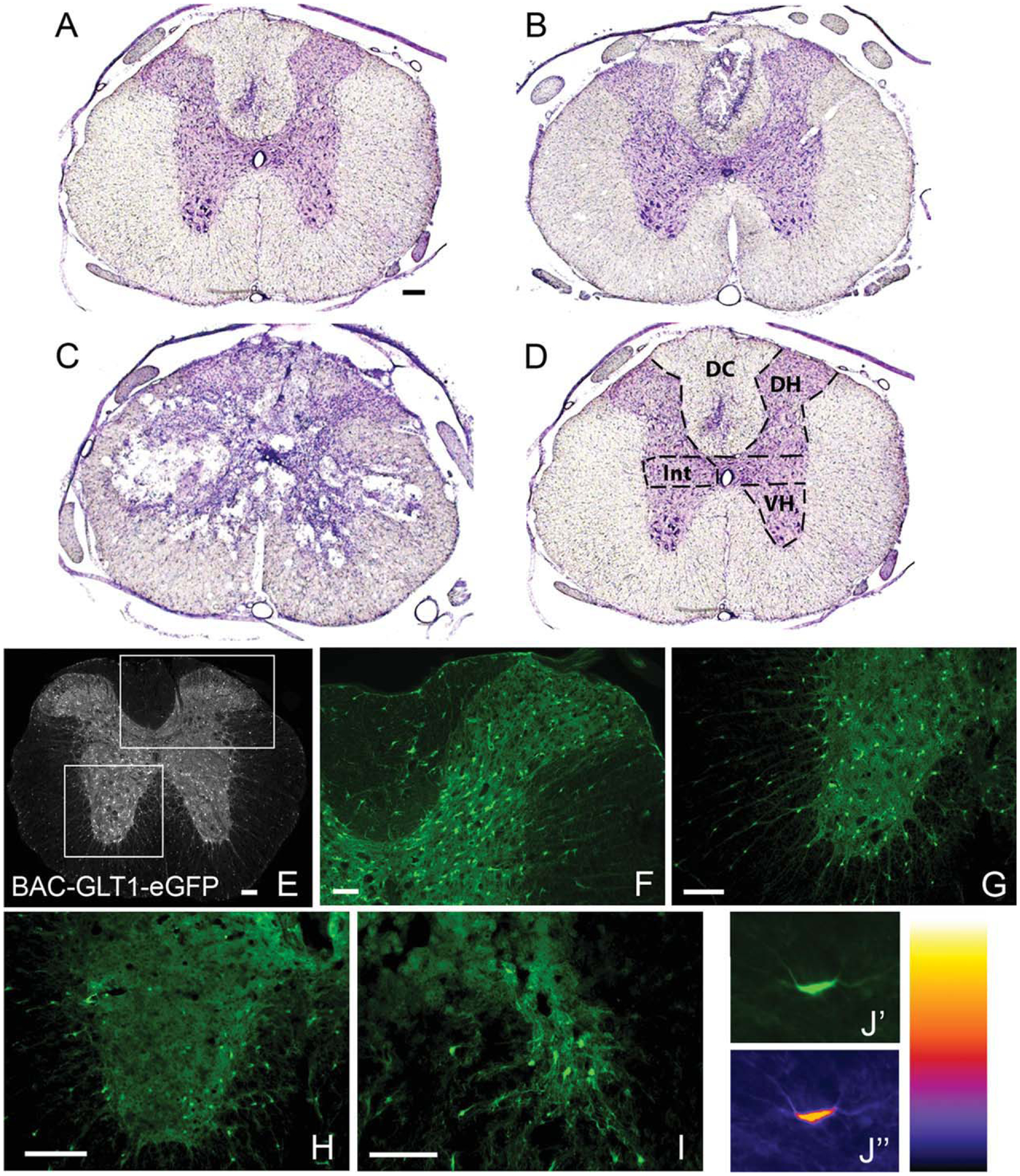
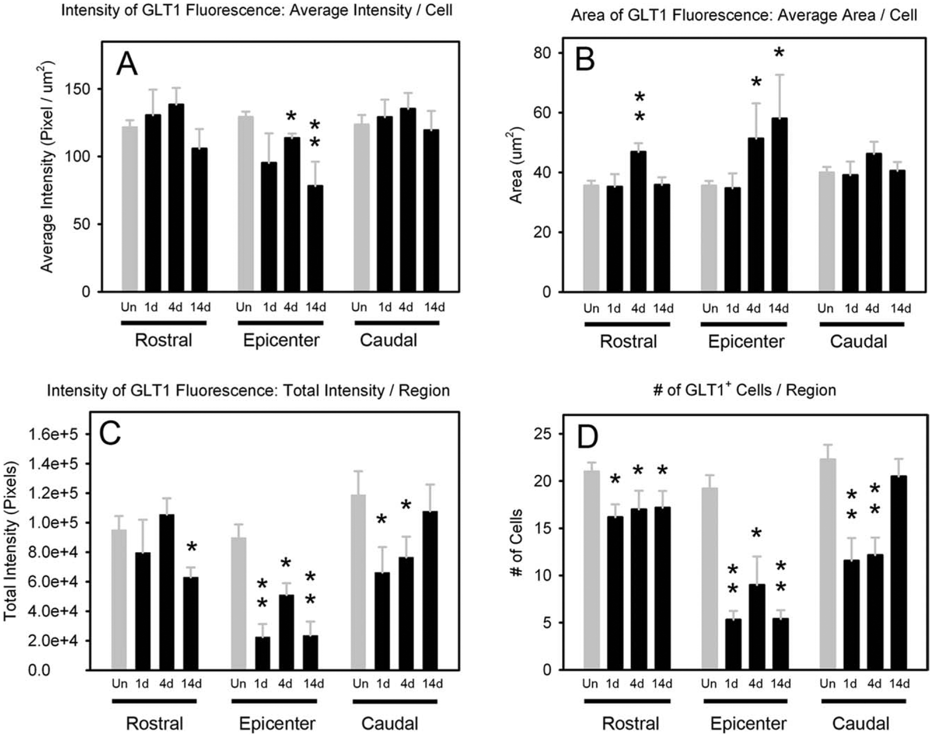
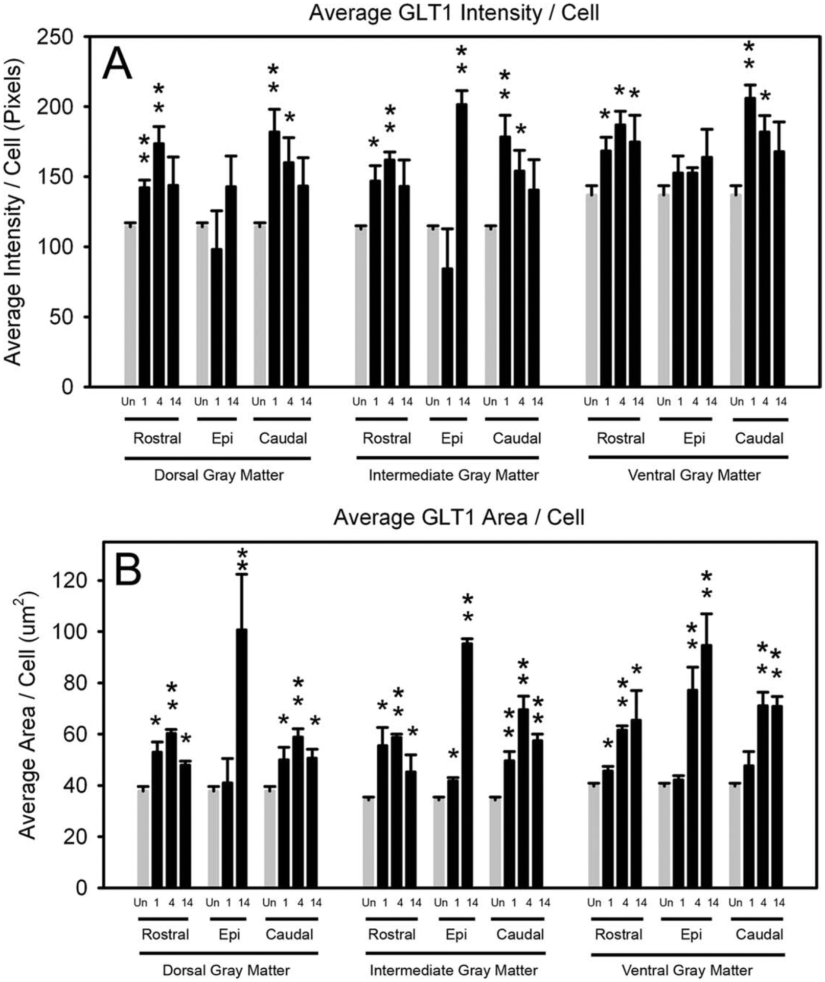
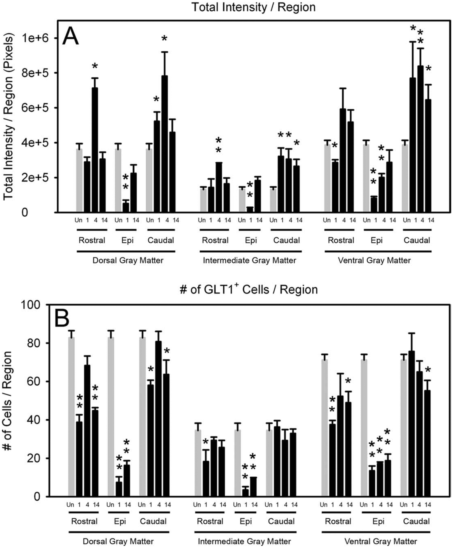
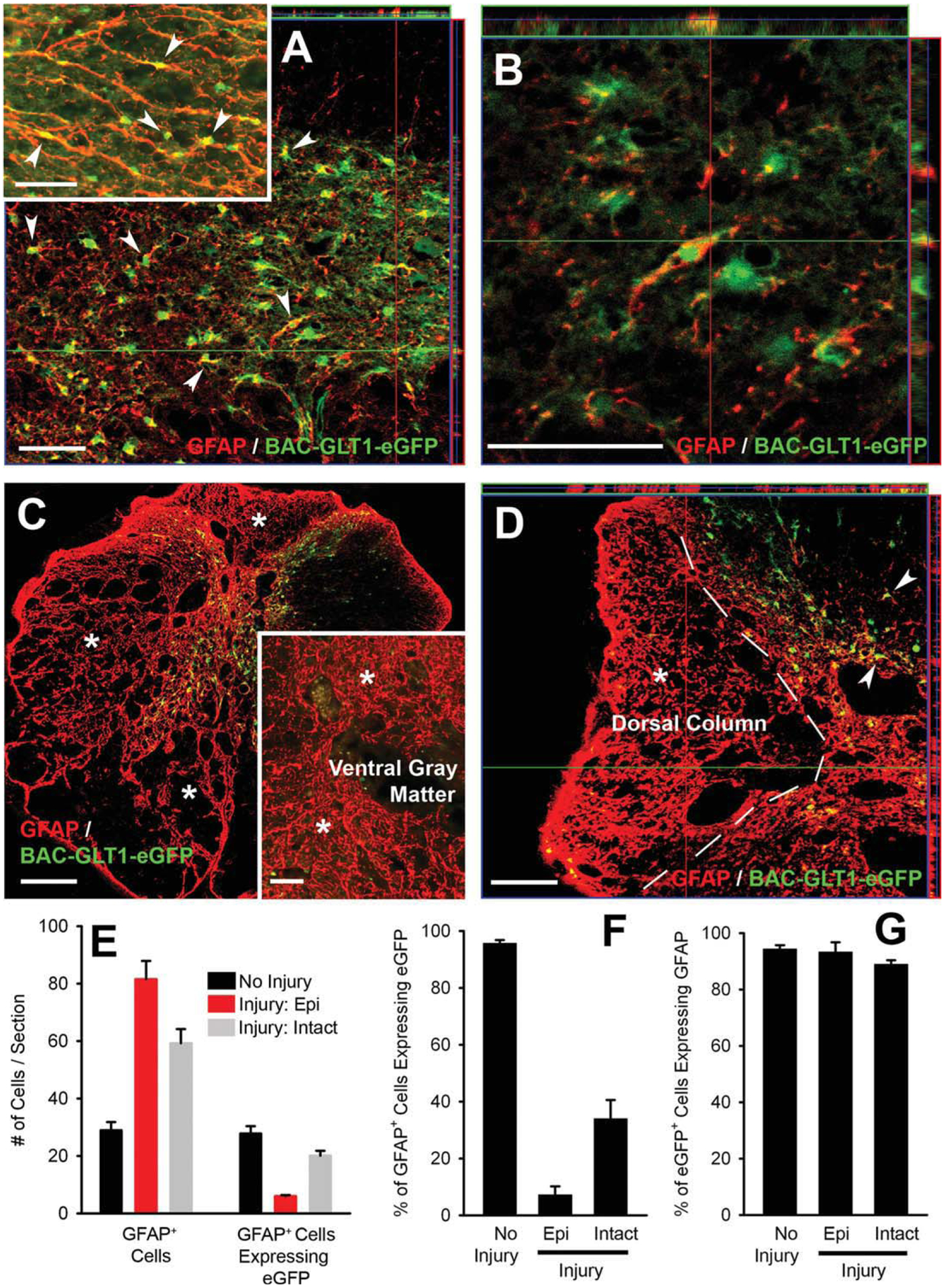
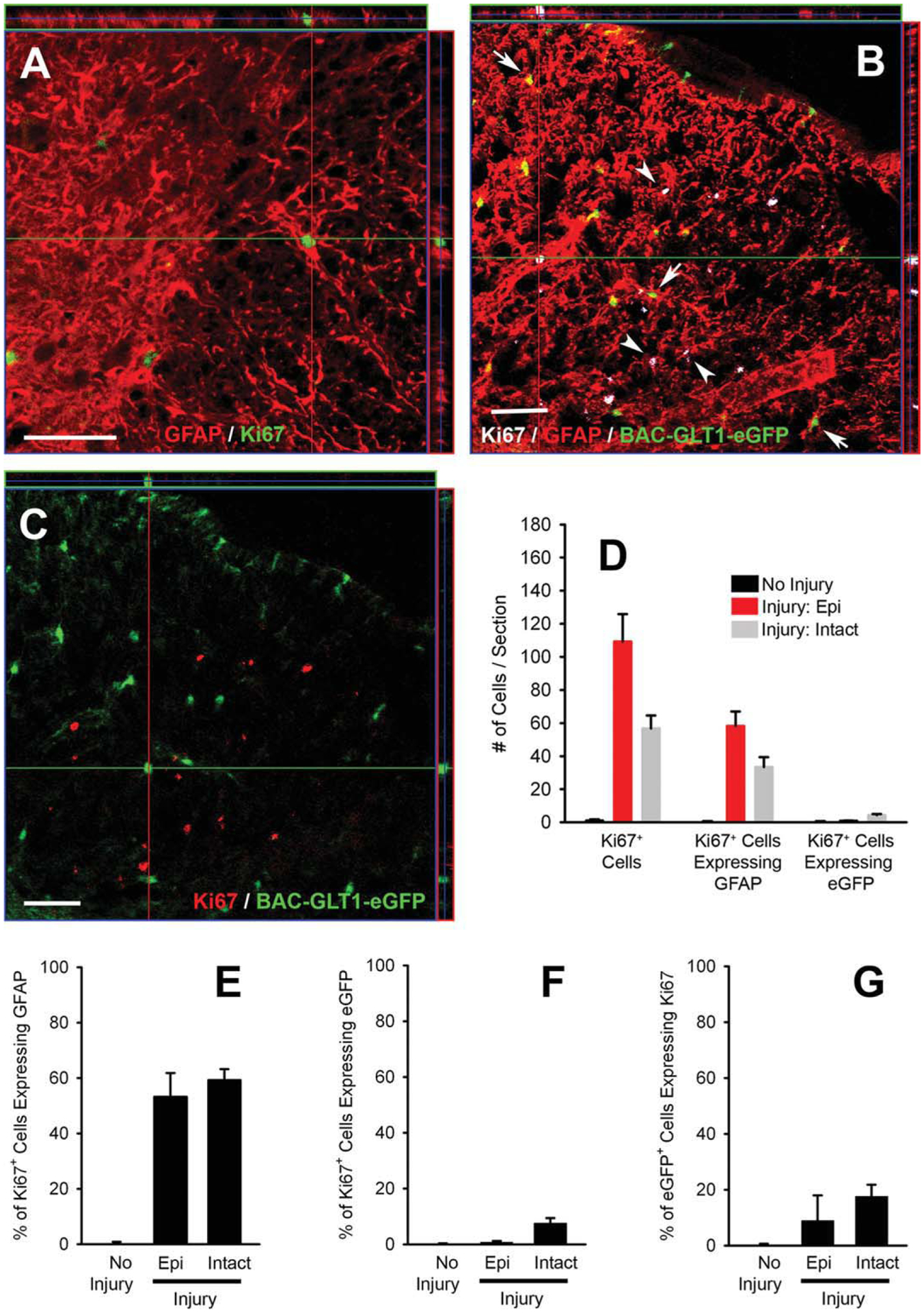
After traumatic spinal cord injury (SCI), there is an opportunity for preserving function by attenuating secondary cell loss. Astrocytes play crucial roles in the adult CNS and are responsible for the vast majority of glutamate buffering, potentially preventing excitotoxic loss of neurons and oligodendrocytes. We examined spatial and temporal changes in gene expression of the major astrocyte glutamate transporter GLT1 following moderate thoracic contusion SCI using transgenic BAC-GLT1-eGFP promoter reporter mice. In dorsal column white matter, total intensity of GLT1-eGFP expression per region was significantly reduced following SCI at both lesion epicenter and at rostral and caudal areas where no tissue loss occurred. This regional decrease in GLT1 expression was due to significant loss of GLT1-eGFP(+) cells, partially accounted for by apoptosis of eGFP(+) /GFAP(+) astrocytes in both white and gray matter. There were also decreased numbers of GLT1-eGFP-expressing cells in multiple gray matter regions following injury; nevertheless, there was sustained or even increased regional GLT1-eGFP expression in gray matter as a result of up-regulation in astrocytes that continued to express GLT1-eGFP. Although there were increased numbers of GFAP(+) cells both at the lesion site and in surrounding intact spinal cord following SCI, the majority of proliferating Ki67(+) /GFAP(+) astrocytes did not express GLT1-eGFP. These findings demonstrate that spatial and temporal alterations in GLT1 expression observed after SCI result from both astrocyte death and gene expression changes in surviving astrocytes. Results also suggest that following SCI a significant portion of astrocytes lacks GLT1 expression, possibly compromising the important role of astrocytes in glutamate homeostasis.


Figures

Fig. 1.
Fig. 1.
Generation of thoracic contusion SCI in BAC-GLT1-eGFP transgenic promoter reporter mice. In cresyl violet-stained sections of thoracic spinal cord from mice that had received a moderate T9 contusion, distinct differences can be observed among intact spinal cord immediately rostral to the injury site (A), the rostral periphery of the lesion site (B), and the center of the injury (C). For BAC-GLT1-eGFP analysis, spinal cord cross-sections were first subdivided into specific anatomical locations (D): dorsal column white matter (DC), dorsal horn gray matter (DH), intermediate gray matter (Int), and ventral horn gray matter (VH). In transgenic BAC-GLT1-eGFP reporter mice, GLT1 promoter activity drives expression of the eGFP reporter (E). In normal, uninjured animals (E–G), GLT1 is found in abundance in gray matter and to a lesser extent in white matter. To analyze GLT1-eGFP expression in astrocytes following SCI (examples, rostral intact cord: H; epicenter: I), individual eGFP+ cells were identified, and the region of fluorescence for each eGFP+ cell was outlined. eGFP average intensity, total intensity, and area for each cell were measured in Metamorph software (J). All images are from mice sacrificed at 14 days postsurgery. Scale bars = 50 μm.
Fig. 2.
Fig. 2.
GLT1-eGFP expression was reduced in dorsal column white matter astrocytes following thoracic contusion in BAC-GLT1-eGFP mice. At 1, 4, and 14 days post-SCI, BAC-GLT1-eGFP mice were analyzed for average intensity (A) and area (B) of eGFP expression per cell (defined as cell expressing eGFP) in dorsal column white matter. The total intensity (C) of eGFP fluorescence per region (sum of the intensity of all individual eGFP-expressing cells in a region) was analyzed following injury. Total numbers of eGFP+ cells were also counted (D). *P < 0.05, **P < 0.001.
Fig. 3.
Fig. 3.
GLT1-eGFP expression in individual gray matter astrocytes increased following thoracic contusion in BAC-GLT1-eGFP mice. At 1, 4, and 14 days after thoracic contusion injury, gray matter at the lesion epicenter and in surrounding intact spinal cord was subdivided into dorsal horn, intermediate and ventral horn, as illustrated in Figure 1D. Each gray matter region was analyzed for average individual cell eGFP fluorescence intensity (A) and average individual eGFP+ cell area (B). *P < 0.05, **P < 0.001.
Fig. 4.
Fig. 4.
Regional GLT1-eGFP expression in gray matter increased following thoracic contusion in BAC-GLT1-eGFP mice. At 1, 4, and 14 days after thoracic contusion injury, each gray matter region was analyzed for combined regional intensities (summation of individual cell intensities of all eGFP+ cells in region; A) and counts of eGFP+ cells (B). *P < 0.05, **P < 0.001.
Fig. 5.
Fig. 5.
GLT1 promoter had limited activity in reactive astrocytes following SCI. In normal uninjured spinal cord, eGFP was expressed only in GFAP+ astrocytes, and nearly all GFAP+ astrocytes expressed eGFP (A,B: image from gray matter; A, inset: image from white matter, F,G). At 4 days following SCI, there was a pronounced increase in GFAP+ astrocytes in dorsal column white matter (E), both at the epicenter (C,D) and in intact spinal cord immediately adjacent to the epicenter of the injury. However, there was also an overall decrease in numbers of eGFP-expressing cells (see Figs. 2–4). Although eGFP expression was still restricted to GFAP+ astrocytes following SCI (D,G), a large proportion of the increased numbers of GFAP+ astrocytes found at the injury site and adjacent spinal cord did not express eGFP (C,D: white matter; C, inset: ventral gray matter, E,F). Areas with large numbers of eGFP astrocytes are denoted by asterisks (C,D,C, inset). Arrowheads denote double-labeled cells in A and D. Scale bars = 50 μm.
Fig. 6.
Fig. 6.
Proliferating astrocytes had little GLT1-eGFP expression following SCI. Proliferating Ki67+/GFAP+ astrocytes were observed throughout the lesioned spinal cord (A,B). In uninjured spinal cord, few proliferating Ki67+ cells were observed (D). At 4 days following injury, there was a large increase in the numbers of proliferating Ki67+ cells in dorsal column white matter, both at the epicenter and adjacent intact spinal cord (D), and approximately half of the proliferating Ki67+ cells were GFAP+ astrocytes (D,E). The vast majority of proliferating astrocytes did not express eGFP (B,C,D,F,G). Arrowheads denote Ki67+/GFAP+/GLT1-eGFP cells. Arrows denote Ki67/GFAP+/GLT1-eGFP+ cells. Scale bars = 50 μm.
Fig. 7.
Fig. 7.
GLT1-eGFP expressing astrocytes underwent apoptosis following SCI. Section from injured spinal cord were triple-labeled for the astrocyte marker GFAP, the apoptosis marker cleaved caspase-3, and eGFP (A). At 4 days following injury, thoracic spinal cord sections from BAC-GLT1-eGFP mice were also double labeled with cleaved caspase-3 and the apoptotic marker TUNEL (B). A control section from injured spinal cord shows positive TUNEL staining (C), as well as negative staining for cleaved caspase-3 in the same section when primary antibody for cleaved capsase-3 was omitted (C′). In uninjured spinal cord, rare apoptotic caspase-3+ cells were observed (D). At 4 days after injury, there was a large increase in the numbers of apoptotic caspase-3+ cells in dorsal column white matter, both at the epicenter and in adjacent intact spinal cord (D). Approximately 20–30% of the apoptotic caspase-3+ cells were GFAP+ astrocytes (D,E). GFAP+/eGFP+ astrocytes that were caspase-3+ were noted both at the epicenter (A) and in adjacent intact spinal cord following SCI but constituted only a small portion of the apoptotic cells (D,F). Nevertheless, approximately 10–20% of eGFP+ cells at 4 days postinjury in the dorsal column white matter were caspase-3+ (D,G). In A, arrows denote caspase-3+/GFAP+/GLT1-eGFP+ cells, and arrowheads denote caspase-3/GFAP+/GLT1-eGFP+ cells. In B, arrows denote TUNEL+/caspase-3+/GLT1-eGFP+ cells. Arrowheads denote TUNEL/caspase-3/GLT1-eGFP+ cells, and asterisks denote apoptotic TUNEL+/caspase-3+ cells that are not GLT1-eGFP+. Scale bars = 50 μm.
Fig. 8.
Fig. 8.
Gray matter changes in astrocyte GLT1 expression, proliferation, and apoptosis following SCI. In ventral gray matter at 4 days postinjury, increased numbers of GFAP+ (A) and Ki67+ (D) cells were noted at both contusion epicenter and adjacent intact spinal cord. A large proportion of these additional GFAP+ cells did not express GLT1-eGFP (A,B), but GLT1-eGFP expression was still mostly restricted to GFAP+ astrocytes (C). Many of the dividing Ki67+ cells were GFAP+ astrocytes (D,E), both at contusion epicenter and in adjacent intact spinal cord. Only a small percentage of dividing Ki67+ cells was GLT1-eGFP+ (F,G). Finally, a significant increase in apoptotic cleaved caspase-3+ cells was observed following SCI, both at contusion epicenter and in adjacent intact spinal cord (H). A proportion of apoptotic cells was GFAP+ (I) and GLT1-eGFP-expressing (J,K) astrocytes.

Similar articles

Cited by

References

    1. Agrawal SK, Fehlings MG. 1997. Role of NMDA and non-NMDA ionotropic glutamate receptors in traumatic spinal cord axonal injury. J Neurosci 17:1055–1063. - PMC - PubMed
    1. Baptiste DC, Fehlings MG. 2006. Pharmacological approaches to repair the injured spinal cord. J Neurotrauma 23:318–334. - PubMed
    1. Basso DM, Fisher LC, Anderson AJ, Jakeman LB, McTigue DM, Popovich PG. 2006. Basso Mouse Scale for locomotion detects differences in recovery after spinal cord injury in five common mouse strains. J Neurotrauma 23:635–659. - PubMed
    1. Cao H, Zhang YQ. 2008. Spinal glial activation contributes to pathological pain states. Neurosci Biobehav Rev 32:972–983. - PubMed
    1. Danbolt NC. 2001. Glutamate uptake. Prog Neurobiol 65:1–105. - PubMed

Publication types

MeSH terms

Substances