. 2011 Jun;79(6):1878-97.
doi: 10.1002/prot.23012. Epub 2011 Apr 12.

Predicted structures of agonist and antagonist bound complexes of adenosine A3 receptor

Affiliations

Predicted structures of agonist and antagonist bound complexes of adenosine A3 receptor

Soo-Kyung Kim et al. Proteins. 2011 Jun.

Abstract

We used the GEnSeMBLE Monte Carlo method to predict ensemble of the 20 best packings (helix rotations and tilts) based on the neutral total energy (E) from a vast number (10 trillion) of potential packings for each of the four subtypes of the adenosine G protein-coupled receptors (GPCRs), which are involved in many cytoprotective functions. We then used the DarwinDock Monte Carlo methods to predict the binding pose for the human A(3) adenosine receptor (hAA(3)R) for subtype selective agonists and antagonists. We found that all four A(3) agonists stabilize the 15th lowest conformation of apo-hAA(3)R while also binding strongly to the 1st and 3rd. In contrast the four A(3) antagonists stabilize the 2nd or 3rd lowest conformation. These results show that different ligands can stabilize different GPCR conformations, which will likely affect function, complicating the design of functionally unique ligands. Interestingly all agonists lead to a trans χ1 angle for W6.48 that experiments on other GPCRs associate with G-protein activation while all 20 apo-AA(3)R conformations have a W6.48 gauche+ χ1 angle associated experimentally with inactive GPCRs for other systems. Thus docking calculations have identified critical ligand-GPCR structures involved with activation. We found that the predicted binding site for selective agonist Cl-IB-MECA to the predicted structure of hAA(3)R shows favorable interactions to three subtype variable residues, I253(6.58), V169(EL2), and Q167(EL2), while the predicted structure for hAA(2A)R shows weakened to the corresponding amino acids: T256(6.58), E169(EL2), and L167(EL2), explaining the observed subtype selectivity.


Figures

Figure 1
Figure 1
Alignments of the four adenosine receptor (AR) subtypes, A1, A2A, A2B, and A3 from the PredicTM method. The predicted TMH regions from PredicTM are displayed in colored boxes (TMH1 in purple, TMH2 in blue, TMH3 in cyan, TMH4 in green, TMH5 in yellow, TMH6 in orange, TMH7 in red), while the TMH region from the x-ray structure of the AA2AR (PDB: 3eml) is underlined. Highly conserved residues in Family A G protein-coupled receptors (GPCRs) are shown in red in TMH 1-6 and white in TMH7. Variable amino acids among the four subtypes in the upper TMH regions are marked with red asterisks and subtype selective residues predicted from the cavity analysis are boxed. The disulfide bridges in yellow were assigned (A1: 80-169, 260-263, A2A: 71-159, 74-146, 77-166, 259-262, A2B: 72-167, 78-171, A3: 83-166) based on the sequence alignment. We use Ballesteros-Weinstein numbering consisting of the TMH helix number followed by residue number relative to the highly conserved residue in the helix, numbered as 50. H-bonding is indicated by arrows, and subtype selective residues are shown in red.
Figure 2
Figure 2
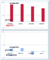
Graphical summary of ensemble docking in Table 3. The relative energies for apo and antagonist or agonist-bound human adenosine A3 receptors (hAA3R) are displayed. We find that all four A3 selective agonists stabilize the 15th lowest conformation of hAA3R while also binding strongly to the 1st and 3rd. In contrast three antagonists stabilize the 2nd lowest conformation of hAA3R while antagonist MRS5127 prefers the 3rd but binding to the 2nd is next best. Experimentally the function of MRS5127 is ambiguous. It appeared to be an antagonist but acted as a partial agonist in cAMP (cyclic Adenosine monophosphate) production in transfected cells with 45% efficacy compared to the full agonist NECA (Adenosine 5’-N-ethyluronamide). All 20 lowest predicted structures for the apo hAA3R have the “toggle switch” W6.48 in the gauche+ χ1 (NH-Cα-Cβ-Cγ) rotamer (tryptophan plane perpendicular to the membrane plane) associated with the inactive G protein-coupled receptor (GPCR) while the lowest predicted structures for the agonist bound state all have the trans χ1 (NH-Cα-Cβ-Cγ) rotamer (tryptophan plane parallel to the membrane plane) associated with the active GPCR. The situation is more ambiguous for the antagonist cases. Here MRS5127 and LJ1251 retain gauche. The cases with this trans rotamer are denoted by filled boxes while the cases with the gauche+ rotamer have open boxes. For cases in which the boxes overlap, the higher energy one was shifted higher to avoid overlapping. These cases are marked with asterisks.
Figure 3
Figure 3
Predicted best structures of selective nucleoside antagonist LJ1251 bound to adenosine A3 receptor (AA3R) (A), selective agonist Cl-IB-MECA bound to adenosine A3 receptor (AA3R) (B) and superimposition of antagonist (blue)/ agonist (red) bound predicted structures (C) without ligands. We predicted the binding to the top 20 predicted structures from the SuperBihelix analysis of the apo-AA3R as shown in Table 3. LJ1251 binds to predicted structure 2 of apo-AA3R while Cl-IB-MECA binds to predicted structure 15 of apo-AA3R The orientation of the W6.48 side chain, shown in yellow, in the agonist-bound hAA3R is perpendicular to the AA3R axis and toward TMH5, while its orientation in the antagonist binding parallel to the AA3R axis allowing it to interacts with the NPxxY motif, just as in the best predicted structure of the apo-AA3R.
Figure 4
Figure 4
The relative difference of total cavity energy depending on the trans vs. gauche+ χ1 (NH-Cα-Cβ-Cγ) rotamer states of W6.48. All top agonist-bound complexes have over ~12 kcal/mol higher E in the gauche+ χ1 form of W6.48 because of steric bumps with 5’-substituents. However, all antagonist-bound predicted structures have similar energies for both conformations. In the case of nucleoside antagonists with 5’-substituents, the 5’-cyclized uronamide in MRS1292 and 5’-N,N-dimethyl uronamide in MRS3771 are < ~3 kcal/mol unfavorable in the gauche+ χ1 form of W6.48. However, MRS5127 and LJ1251, which were truncated at the 5’-position, are more stable in the gauche+ χ1 conformation of W6.48.
Figure 5
Figure 5
Molecular dynamics simulation of (A) antagonist LJ1251 in yellow and (B) agonist Cl-IB-MECA in orange bound human adenosine A3 receptor (hAA3R) in explicit lipid and water. Bottom) χ1 torsion angle (NH-Cα-Cβ-Cγ) variation of W2436.48 in green through 10 ns simulations. Ligand at the beginning is represented by the stick model and ligand at 10ns is shown in vdW (van der Waals) drawing mode of the VMD (Visual Molecular Dynamics) program.
Figure 6
Figure 6
Schematic of the predicted binding sites for adenosine receptor agonists based on the cavity binding energies. Left: adenosine at hAA2AR; Right: Cl-IB-MECA at hAA3R
Figure 7
Figure 7
Predicted binding sites (A) adenosine to the adenosine A2A receptor (AA2AR), (B) adenosine to the adenosine A3 receptor (AA3R), (C) A3 selective agonist Cl-IB-MECA at AA2AR, (D) A3 selective agonist Cl-IB-MECA at AA3Rs. The high selectivity of Cl-IB-MECA for AA3R relative to AA2AR is due to the salt-bridge between E169 and H264 (red circle in (C)) in AA3R. In AA2AR we have V169 but there is no residue corresponding to H264 because of the gap in AA2AR. H-bonding is indicated by red dots, and subtype selective residues are shown in red.
Chart 1
Chart 1
Structures of four agonists (Ag) and four antagonists (Ant) exhibiting selective binding to the adenosine A3 receptor with respect to A2a. Binding affinities (Ki in nM) are shown for A3 with relative efficacies in parenthesis compared to the endogenous adenosine.

Similar articles

Cited by

References

    1. Jacobson KA, Gao ZG. Adenosine receptors as therapeutic targets. Nat Rev Drug Discov. 2006;5:247–264. - PMC - PubMed
    1. Jacobson KA, Costanzi S, Kim S-K, Rho E, Joshi BV, Tchilibon S, Duong HT, Gao Z-G. Nucleosides, Action of nucleosides and nucleotides at 7 transmembrane-spanning receptors. Nucleotides, and Nucleic Acids. 2006;25:1425–1436. - PMC - PubMed
    1. Vu CB, Shields P, Peng B, Kumaravel G, Jin X, Phadke D, Wang J, Engber T, Ayyub E, Petter RC. Triamino derivatives of triazolotriazine and triazolopyrimidine as adenosine A2A receptor antagonists. Bioorg Med Chem Lett. 2004;14:4835–4838. - PubMed
    1. Tracey WR, Magee WP, Oleynek JJ, Hill RJ, Smith AH, Flynn DM, Knight DR. Novel N6-substituted adenosine 5’-N-methyluronamides with high selectivity for human adenosine A3 receptors reduce ischemic myocardial injury. Am J Physiol Heart Circ Physiol. 2003;285:H2780–2787. - PubMed
    1. Shneyvais V, Mamedova L, Zinman T, Jacobson KA, Shainberg A. Activation of A3 adenosine receptor protects against doxorubicin-induced cardiotoxicity. J Mol Cell Cardiol. 2001;33:1249–1261. - PMC - PubMed

Publication types

MeSH terms

Substances

LinkOut - more resources