

Neuron-like differentiation of bone marrow-derived mesenchymal stem cells
- PMID: 21488182
- PMCID: PMC3101055
- DOI: 10.3349/ymj.2011.52.3.401
Neuron-like differentiation of bone marrow-derived mesenchymal stem cells
Abstract
Purpose: Mesenchymal stem cells (MSCs) are multipotent and give rise to distinctly differentiated cells from all three germ layers. Neuronal differentiation of MSC has great potential for cellular therapy. We examined whether the cluster of mechanically made, not neurosphere, could be differentiated into neuron-like cells by growth factors, such as epidermal growth factor (EGF), hepatocyte growth factor (HGF), and vascular endothelial growth factor (VEGF).
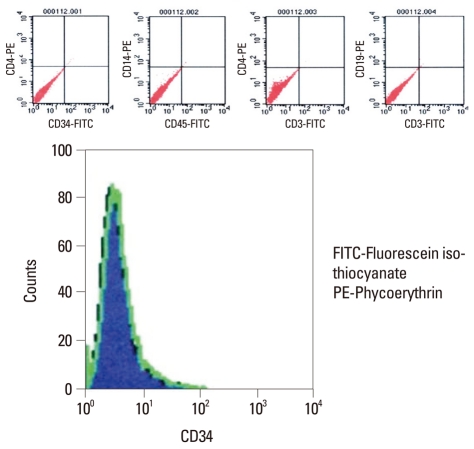
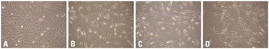
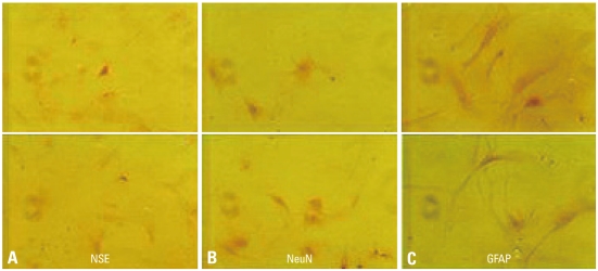
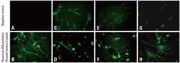
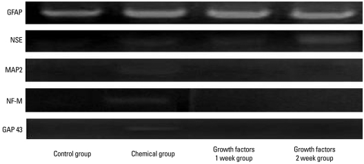
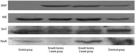
Materials and methods: BMSCs grown confluent were mechanically separated with cell scrapers and masses of separated cells were cultured to form cluster BMSCs. As described here cluster of BMSCs were differentiated into neuron-like cells by EGF, HGF, and VEGF. Differentiated cells were analyzed by means of phase-contrast inverted microscopy, reverse transcriptase-polymerase chain reaction (RT-PCR), immunofluorescence, and immunocytochemistry to identify the expression of neural specific markers.
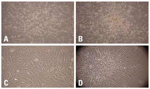
Results: For the group with growth factors, the shapes of neuron-like cells was observable a week later, and two weeks later, most cells were similar in shape to neuron-like cells. Particularly, in the group with chemical addition, various shapes of filament structures were seen among the cells. These culture conditions induced MSCs to exhibit a neural cell phenotype, expressing several neuro-glial specific markers.
Conclusion: bone marrow-derived mesenchymal stem cells (BMSCs) could be easily induced to form clusters using mechanical scraping, not neurospheres, which in turn could differentiate further into neuron-like cells and might open an attractive possibility for clinical cell therapy for neurodegenerative diseases. In the future, we consider that neuron-like cells differentiated from clusters of BMSCs are needed to be compared and analyzed on a physiological and molecular biological level with preexisting neuronal cells, and studies on the possibility of their transplantation and differentiation capability in animal models are further required.
Conflict of interest statement
The authors have no financial conflicts of interest.
Figures
References
-
- Owen M. Lineage of osteogenic cells and their relationship to the stromal system. In: Peck WA, editor. Bone and mineral research. Amsterdam: Elsevier; 1985. pp. 1–25.
-
- Pittenger MF, Mackay AM, Beck SC, Jaiswal RK, Douglas R, Mosca JD, et al. Multilineage potential of adult human mesenchymal stem cells. Science. 1999;284:143–147. - PubMed
-
- Tremain N, Korkko J, Ibberson D, Kopen GC, DiGirolamo C, Phinney DG. MicroSAGE analysis of 2,353 expressed genes in a single cell-derived colony of undifferentiated human mesenchymal stem cells reveals mRNAs of multiple cell lineages. Stem Cells. 2001;19:408–418. - PubMed
-
- Le Blanc K, Pittenger M. Mesenchymal stem cells: progress toward promise. Cytotherapy. 2005;7:36–45. - PubMed
-
- Lee RH, Kim B, Choi I, Kim H, Choi HS, Suh K, et al. Characterization and expression analysis of mesenchymal stem cells from human bone marrow and adipose tissue. Cell Physiol Biochem. 2004;14:311–324. - PubMed
MeSH terms
Substances
LinkOut - more resources
Full Text Sources
Other Literature Sources
Research Materials
