. 2011 Jun;28(6):871-88.
doi: 10.1089/neu.2010.1586. Epub 2011 Jun 1.

Acute serum hormone levels: characterization and prognosis after severe traumatic brain injury

Affiliations

Acute serum hormone levels: characterization and prognosis after severe traumatic brain injury

Amy K Wagner et al. J Neurotrauma. 2011 Jun.

Abstract

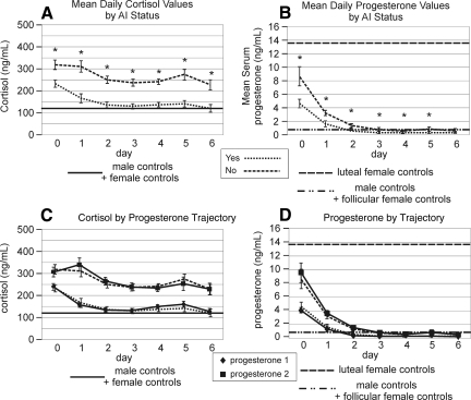
Experimental traumatic brain injury (TBI) studies report the neuroprotective effects of female sex steroids on multiple mechanisms of injury, with the clinical assumption that women have hormonally mediated neuroprotection because of the endogenous presence of these hormones. Other literature indicates that testosterone may exacerbate injury. Further, stress hormone abnormalities that accompany critical illness may both amplify or blunt sex steroid levels. To better understand the role of sex steroid exposure in mediating TBI, we 1) characterized temporal profiles of serum gonadal and stress hormones in a population with severe TBI during the acute phases of their injury; and 2) used a biological systems approach to evaluate these hormones as biomarkers predicting global outcome. The study population was 117 adults (28 women; 89 men) with severe TBI. Serum samples (n=536) were collected for 7 days post-TBI for cortisol, progesterone, testosterone, estradiol, luteinizing hormone (LH), and follicle-stimulating hormone (FSH). Hormone data were linked with clinical data, including acute care mortality and Glasgow Outcome Scale (GOS) scores at 6 months. Hormone levels after TBI were compared to those in healthy controls (n=14). Group based trajectory analysis (TRAJ) was used to develop temporal hormone profiles that delineate distinct subpopulations in the cohort. Structural equations models were used to determine inter-relationships between hormones and outcomes within a multivariate model. Compared to controls, acute serum hormone levels were significantly altered after severe TBI. Changes in the post-TBI adrenal response and peripheral aromatization influenced hormone TRAJ profiles and contributed to the abnormalities, including increased estradiol in men and increased testosterone in women. In addition to older age and greater injury severity, increased estradiol and testosterone levels over time were associated with increased mortality and worse global outcome for both men and women. These findings represent a paradigm shift when thinking about the role of sex steroids in neuroprotection clinically after TBI.


Figures

FIG. 1.
FIG. 1.
(A) Schematic of peripheral steroidogenesis highlighting progesterone, cortisol, estradiol, and testosterone. The adrenal gland can produce each of these hormones, and peripheral conversion of testosterone to estradiol can occur in cell types including adipocytes, osteoblasts, and fibroblasts. (B) Theoretical multivariate model for SEQM modeling that incorporates biologically relevant relationships between hormones for prediction of outcome. Theoretical hormone prognostic model: direct effects from hormones and demographic variables on outcome are represented by arrows. Indirect (mediated) effects from progesterone on outcome through other hormones are represented by *. SEQM, structural equations modeling.
FIG. 2.
FIG. 2.
Daily hormone levels by sex over the first 6 days after severe TBI compared to those of healthy controls. (A) Mean serum cortisol levels. There were no significant sex differences in cortisol levels over the time course. (B) Mean serum progesterone levels. There were no significant sex differences levels over the time course. (C) Mean serum testosterone levels. Female TBI subjects had testosterone levels higher than controls, and male values were higher than female values for days 0–2 (p<0.001 all comparisons). (D) Mean serum estradiol levels. Estradiol values for men were elevated compared to controls on days 0–2, and estradiol levels for women were significantly higher than for men only on day 0 (p<0.024 all comparisons). (E) Mean serum FSH levels. Values were significantly higher for women compared to those for men on days 0–5 (p<0.004 all comparisons). (F) Mean serum LH levels. Values were significantly higher for women compared to men on days 0–1 and 4 (p<0.001 all comparisons).
FIG. 3.
FIG. 3.
(A) Trajectory groups for profiles over time and percent membership for each trajectory group for serum cortisol using ranked data. The y-axis represents the relative rank. TRAJ identified three groups for cortisol: group 1=decliner group, group 2=low group, 3=high group. (B) Mean serum cortisol concentrations over time for each cortisol trajectory group compared to those of healthy controls. There were significant differences in TRAJ group cortisol levels on days 0–6 (p<0.001 all comparisons).
FIG. 4.
FIG. 4.
(A) Trajectory group profiles over time and percent membership for each TRAJ group for serum progesterone using ranked data. The y-axis represents the relative rank. TRAJ identified two groups for progesterone: 1=low group, 2=high group. (B) Mean serum progesterone concentrations over time for each progesterone trajectory group compared to those of healthy controls. There were significant differences in TRAJ group progesterone levels on days 0–6 (p<0.001 all comparisons).
FIG. 5.
FIG. 5.
(A) TRAJ groups for profiles over time and percent membership for each TRAJ group for serum testosterone using ranked data. The y-axis represents the relative rank. TRAJ identified three groups for testosterone. 1=low group, 2=decliner group, 3=high group. (B) Mean serum testosterone concentrations over time for each testosterone TRAJ group compared to those of healthy controls. There were significant differences in TRAJ group testosterone levels on days 0–5 (p<0.001 all comparisons).
FIG. 6.
FIG. 6.
(A) TRAJ groups for profiles over time and percent membership for each TRAJ group for serum estradiol using ranked data. The y-axis represents the relative rank. TRAJ identified four groups for estradiol: 1=low group, 2=riser group, 3=middle group, 4=high group. (B) Mean serum estradiol concentrations over time for each estradiol TRAJ group compared to those of healthy controls. There were significant differences in TRAJ group estradiol levels on days 0–5 (p<0.001 all comparisons).
FIG. 7.
FIG. 7.
Daily serum hormone levels by progesterone TRAJ and compared to those of healthy controls. (B) Serum progesterone levels. Hormone levels for each TRAJ group were significantly different from each other on days 0–6 post-TBI (p<0.001 all comparisons. (A) Serum cortisol levels. Hormone levels for each TRAJ group were significantly different from each other on days 0–6 post-TBI (p<0.025 all comparisons. (C) Serum testosterone levels. Hormone levels for each TRAJ group were not significantly different from each other on any day. (D) Serum estradiol levels. Hormone levels for each TRAJ group were significantly different from each other on days 3–4 post-TBI (p<0.018 both comparisons). (E) Serum FSH levels. Hormone levels for each TRAJ group were significantly different from each other on days 0–3 post-TBI (p<0.039 all comparisons). (F) Serum LH levels. Hormone levels for each TRAJ group were significantly different from each other on days 0–1 post-TBI (p<0.002 both comparisons).
FIG. 8.
FIG. 8.
Daily serum profiles compared to those of healthy controls. (B) Progesterone levels by AI status. Subjects with AI have significantly lower progesterone levels for days 0–6 (p<0.001 all comparisons). (A) Cortisol levels by AI status. Subjects with AI had significantly lower cortisol levels for days 0–6 (p<0.001 all comparisons). (C) Progesterone levels by AI status with progesterone levels by progesterone TRAJ superimposed to demonstrate similarity of progesterone profiles over time. (D) Cortisol levels by AI status with cortisol levels by progesterone TRAJ superimposed to demonstrate similarity of cortisol profiles over time. AI, adrenal insufficiency.
FIG. 9.
FIG. 9.
SEQM with acute mortality as outcome. Standardized coefficients are shown for direct comparison of hormonal effects on outcome. Mediated effects of progesterone and their 95% bias-corrected bootstrap confidence intervals are shown in the table. Significant direct effects on outcome: age (p=0.003), GCS (p=0.002), estradiol (p=0.026), and testosterone (p=0.033). Progesterone has significant direct effect on estradiol (p<0.001) and cortisol (p<0.001), and a trend on testosterone (p=0.072). *Progesterone also has a significant indirect (mediated) effect on outcome through estradiol (p=0.049). SEQM. structural equations modeling.
FIG. 10.
FIG. 10.
SEQM with GOS at 6 month as outcome. Standardized coefficients are shown for direct comparison of hormonal effects on outcome. Mediated effects of progesterone and their 95% bias-corrected bootstrap confidence intervals are shown in the table. Significant direct effects on outcome: age (p=0.002), GCS (p=0.001), and estradiol (p=0.021). Testosterone trends toward significance (p=0.163). Progesterone has significant direct effect on estradiol (p<0.001) and cortisol (p<0.001), and a trend on testosterone (p=0.072). *Progesterone also has significant indirect (mediated) effect on outcome via estradiol (p=0.045). SEQM: structural equations modeling; GOS: Glasgow Outcome Scale.

References

    1. Agha A. Rogers B. Sherlock M. O'Kelly P. Tormey W. Phillips J. Thompson C.J. Anterior pituitary dysfunction in survivors of traumatic brain injury. J. Clin. Endocrinol. Metab. 2004;89:4929–4936. - PubMed
    1. Azcoitia I. Sierra A. Veiga S. Garcia–Segura L.M. Aromatase expression by reactive astroglia is neuroprotective. Ann. N. Y. Acad. Sci. 2003;1007:298–305. - PubMed
    1. Bavisetty S. McArthur D.L. Dusick J.R. Wang C. Cohan P. Boscardin W.J. Swerdloff R. Levin H. Chang D.J. Muizelaar J.P. Kelly D.F. Chronic hypopituitarism after traumatic brain injury: risk assessment and relationship to outcome. Neurosurgery. 2008;62:1080–1093. - PubMed
    1. Beale E. Zhu J. Belzberg H. Changes in serum cortisol with age in critically ill patients. Gerontology. 2002;48:84–92. - PubMed
    1. Behan L.A. Phillips J. Thompson C.J. Agha A. Neuroendocrine disorders after traumatic brain injury. J. Neurol. Neurosurg. Psychiatr. 2008;79:753–759. - PubMed

Publication types

Substances