

Thrombomodulin phenotype of a distinct monocyte subtype is an independent prognostic marker for disseminated intravascular coagulation
- PMID: 21489300
- PMCID: PMC3219396
- DOI: 10.1186/cc10139
Thrombomodulin phenotype of a distinct monocyte subtype is an independent prognostic marker for disseminated intravascular coagulation
Abstract
Introduction: Thrombomodulin, which is expressed solely on monocytes, along with tissue factor (TF), takes part in coagulation and inflammation. Circulating blood monocytes can be divided into 3 major subtypes on the basis of their receptor phenotype: classical (CD14brightCD16negative, CMs), inflammatory (CD14brightCD16positive; IMs), and dendritic cell-like (CD14dimCD16positive DMs). Monocyte subtype is strongly regulated, and the balance may influence the clinical outcomes of disseminated intravascular coagulation (DIC). Therefore, we investigated the phenotypic difference in thrombomodulin and TF expression between different monocyte subtypes in coagulopathy severity and prognosis in patients suspected of having DIC.
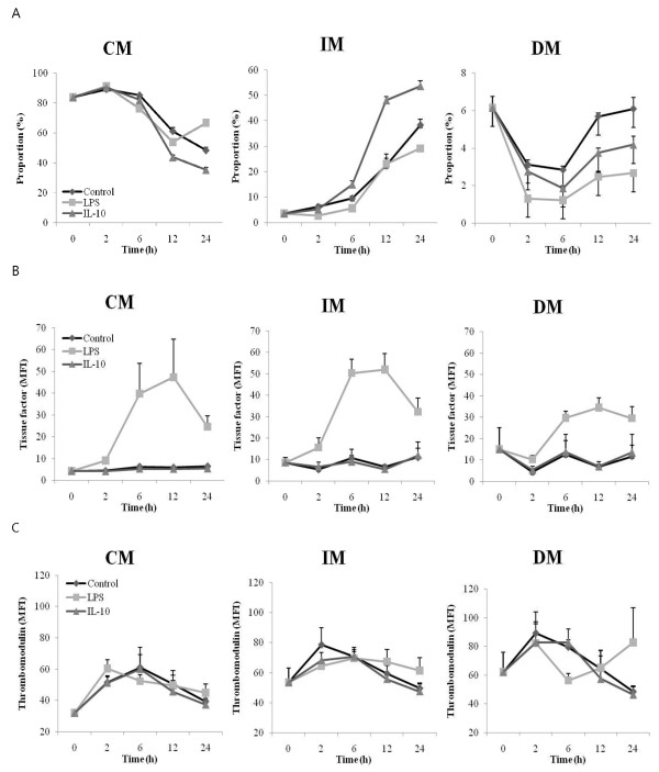
Methods: In total, 98 patients suspected of having DIC were enrolled. The subtypes of circulating monocytes were identified using CD14 and CD16 and the thrombomodulin and TF expression in each subtype, expressed as mean fluorescence intensity, was measured by flow cytometry. Plasma level of tissue factor was measured by ELISA. In cultures of microbead-selected, CD14-positive peripheral monocytes, lipopolysaccharide (LPS)- or interleukin-10-induced expression profiles were analyzed, using flow cytometry.
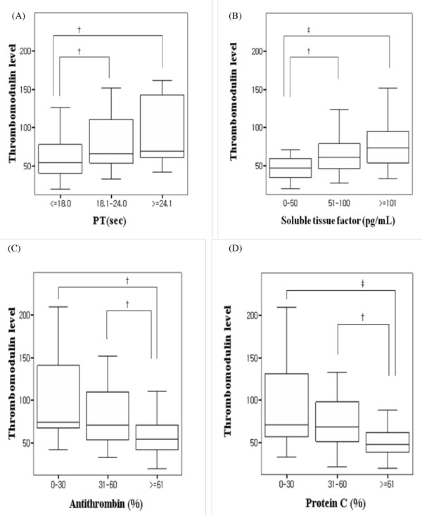
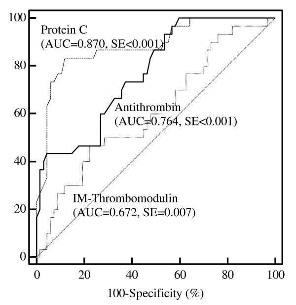
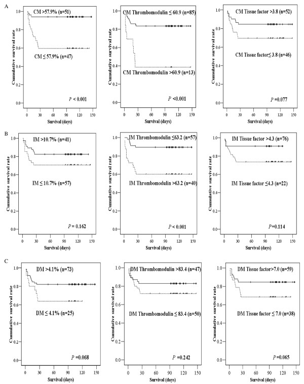
Results: The proportion of monocyte subtypes did not significantly differ between the overt and non-overt DIC groups. The IM thrombomodulin expression level was prominent in the overt DIC group and was well correlated with other coagulation markers. Of note, IM thrombomodulin expression was found to be an independent prognostic marker in multivariate Cox regression analysis. In addition, in vitro culture of peripheral monocytes showed that LPS stimulation upregulated thrombomodulin expression and TF expression in distinct populations of monocytes.
Conclusions: These findings suggest that the IM thrombomodulin phenotype is a potential independent prognostic marker for DIC, and that thrombomodulin-induced upregulation of monocytes is a vestige of the physiological defense mechanism against hypercoagulopathy.
Figures
Similar articles
-
Decreased thrombomodulin mRNA expression on peripheral monocytes in disseminated intravascular coagulation patients relates to poor outcomes: the ex vivo effects of lipopolysaccharide and thrombin on monocyte thrombomodulin and CD14 mRNA.Thromb Res. 2013 Sep;132(3):392-7. doi: 10.1016/j.thromres.2013.07.025. Epub 2013 Aug 3. Thromb Res. 2013. PMID: 23954259
-
Circulating hepatocyte growth factor as an independent prognostic factor of disseminated intravascular coagulation.Thromb Res. 2010 Jun;125(6):e285-93. doi: 10.1016/j.thromres.2010.01.046. Epub 2010 Feb 24. Thromb Res. 2010. PMID: 20185166
-
Plasma level of stromal derived factor-1 (SDF-1) is increased in disseminated intravascular coagulation patients who have poor outcomes: in vitro effect of SDF-1 on coagulopathy.Thromb Res. 2007;120(4):559-66. doi: 10.1016/j.thromres.2006.11.011. Epub 2007 Jan 18. Thromb Res. 2007. PMID: 17239427
-
Tissue factor in trauma and organ dysfunction.Semin Thromb Hemost. 2006 Feb;32(1):48-53. doi: 10.1055/s-2006-933340. Semin Thromb Hemost. 2006. PMID: 16479462 Review.
-
Sepsis and disseminated intravascular coagulation.J Intensive Care. 2016 Mar 23;4:23. doi: 10.1186/s40560-016-0149-0. eCollection 2016. J Intensive Care. 2016. PMID: 27011792 Free PMC article. Review.
Cited by
-
Leukocyte Count and Intracerebral Hemorrhage Expansion.Stroke. 2016 Jun;47(6):1473-8. doi: 10.1161/STROKEAHA.116.013176. Epub 2016 Apr 21. Stroke. 2016. PMID: 27103016 Free PMC article.
-
Association Between Eosinophilic Leukocyte Count and Hematoma Expansion in Acute Spontaneous Intracerebral Hemorrhage.Front Neurol. 2019 Oct 31;10:1164. doi: 10.3389/fneur.2019.01164. eCollection 2019. Front Neurol. 2019. PMID: 31736868 Free PMC article.
-
The immunoregulatory role of monocytes and thrombomodulin in myelodysplastic neoplasms.Front Oncol. 2024 Jul 26;14:1414102. doi: 10.3389/fonc.2024.1414102. eCollection 2024. Front Oncol. 2024. PMID: 39132505 Free PMC article. Review.
References
-
- Persson E, Olsen O. Current status on tissue factor activation of factor VIIa. Thromb Res. 2010;125:S11–S12. - PubMed
Publication types
MeSH terms
Substances
LinkOut - more resources
Full Text Sources
Other Literature Sources
Research Materials
Miscellaneous
