Review
. 2011 Jun 15;69(12):e89-99.
doi: 10.1016/j.biopsych.2011.01.027. Epub 2011 Apr 13.

Catecholamine influences on dorsolateral prefrontal cortical networks

Affiliations
Review

Catecholamine influences on dorsolateral prefrontal cortical networks

Amy F T Arnsten. Biol Psychiatry. .

Abstract

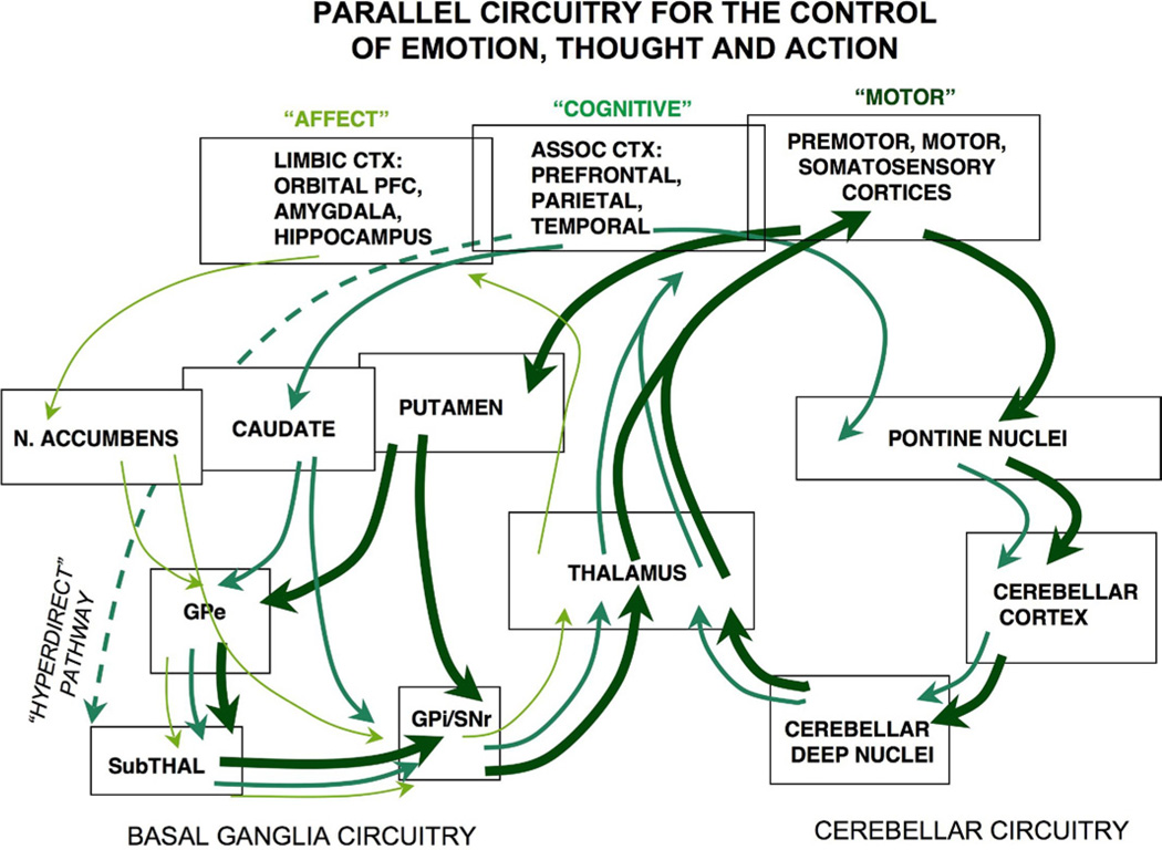
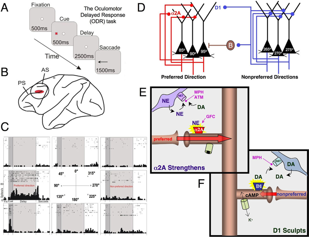
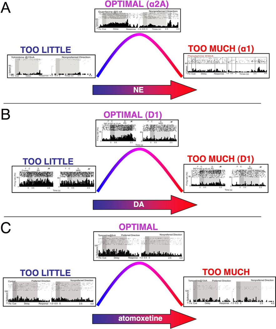
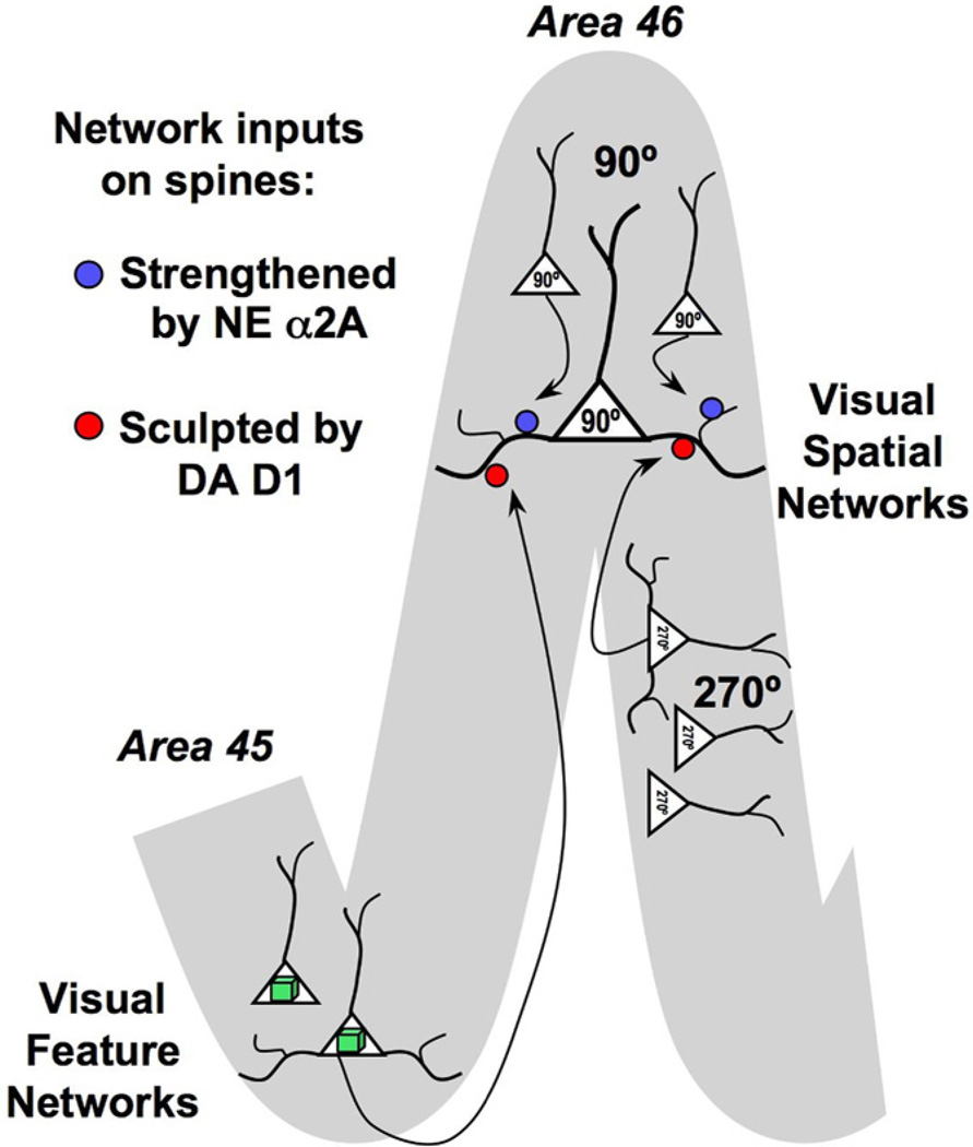
The symptoms of attention-deficit/hyperactivity disorder (ADHD) involve impairments in prefrontal cortical top-down regulation of attention and behavior. All current pharmacological treatments for ADHD facilitate catecholamine transmission, and basic research suggests that these compounds have prominent actions in the prefrontal cortex (PFC). The dorsolateral PFC is especially sensitive to levels of norepinephrine and dopamine, whereby either too little or too much markedly impairs PFC function. Recent physiological studies have shown that norepinephrine strengthens PFC network connectivity and maintains persistent firing during a working memory task through stimulation of postsynaptic α(2A)-adrenoceptors on PFC neurons. Conversely, dopamine acts at D1 receptors to narrow spatial tuning, sculpting network inputs to decrease noise (i.e., stabilization of the representation). The stimulant medications and atomoxetine appear to enhance PFC function by indirectly increasing these catecholamine actions through blockade of norepinephrine and/or dopamine transporters. In contrast, guanfacine mimics the enhancing effects of norepinephrine at postsynaptic α(2A)-receptors in the PFC, strengthening network connectivity. Stronger PFC regulation of attention, behavior, and emotion likely contributes to the therapeutic effects of these medications for the treatment of ADHD.


Figures

Figure 1
Figure 1
Parallel basal ganglia and cerebellar loops for the control of behavior, thought, and emotion. The pioneering work of Middleton and Strick (9) has revealed parallel pathways regulating motor response, cognition, and emotion. One set of pathways projects from cortex through striatal structures in the basal ganglia to focus back on prefrontal cortex for the selection and planning of motor, cognitive, and emotional habits. A second set of pathways project through the pontine nuclei to the cerebellum; indeed, the majority of the cerebellar cortex in primates receives projections from nonmotor cortices. Please note that some prefrontal cortex regions also project directly to the subthalamic nucleus for rapid stopping of behavioral responses (not shown). Alterations in these pathways may often contribute to the symptoms of attention-deficit/hyperactivity disorder. ASSOC, association; CTX, cortex; GPe, globus pallidus external segment; GPi, globus pallidus internal segment; N. ACCUMBENS, nucleus accumbens; SNr, substantia nigra pars reticulata; SubTHAL, subthalamic nucleus.
Figure 2
Figure 2
Spatial working memory networks in the dorsolateral prefrontal cortex (PFC). (A) The oculomotor delayed response task is a spatial working memory task that is used to probe the physiological profiles of PFC neurons. The subject must remember the spatial position of the most recent cue over a delay period of several seconds and then indicate that position with a saccade to the memorized location. (B) The region of the monkey dorsolateral PFC (Walker’s area 46) where neurons show persistent, spatially tuned firing during the oculomotor delayed response task. (C) An example of a PFC neuron that shows persistent firing during the delay period if the cue had occurred at 90° (the preferred direction for this neuron) but not at other spatial locations. A nonpreferred direction opposite to the preferred direction is also labeled for reference to Figure 3 in which only the preferred and one nonpreferred direction are shown. Rasters show the firing of the neuron over seven trials at each spatial position. (D) A schematic drawing of the PFC microcircuits underlying spatial working memory as described by Goldman-Rakic (23). Layer III pyramidal cells (black) receive highly processed spatial information from parietal association cortices (not shown). Pyramidal cells with similar spatial characteristics engage in recurrent excitation to maintain persistent activity over the delay period (note that the subcellular localization of these excitatory connections is not currently known; they could be on the apical and/or basal dendrites). Gamma-aminobutyric acidergic interneurons help to spatially tune neurons through lateral inhibition; one of these is labeled as B (basket cell). Network synaptic inputs from isodirectional inputs (neurons with the same tuning profile) are shown in red, while inputs form cross-directional microcircuits (neurons with different tuning characteristics) are shown in blue. (E) A working model of norepinephrine (NE) actions at α2A receptors on PFC dendritic spines. Stimulation of α2A receptors inhibits cyclic adenosine monophosphate signaling, which closes nearby potassium channels and strengthens the efficacy of network connections onto the spine. The physiological data suggest that these actions occur on spines receiving preferred network inputs (Wang et al. [57]). Many of the drugs approved to treat attention-deficit/hyperactivity disorder block the NE transporter and increase NE (and dopamine [DA]) availability, e.g., medications such as methylphenidate and atomoxetine. In contrast, guanfacine mimics NE actions by directly stimulating the α2A receptor. (F) Physiological studies in monkeys performing working memory tasks indicate that an optimal level of DA D1 receptor stimulation weakens neuronal firing for nonpreferred inputs, thus enhancing the spatial tuning of the neuron (Vijayraghavan et al. [109]). Physiological recordings indicate that these sculpting actions involve increased cyclic adenosine monophosphate signaling, likely via opening of potassium channels. Stimulants such as methylphenidate block the DA transporter to increase DA availability; blockade of the NE transporter similarly increases DA availability in the PFC (see text). AS, arcuate sulcus; ATM, atomoxetine; B, basket cell; DA, dopamine; DAT, dopamine transporter; GFC, guanfacine; K+, potassium; MPH, methylphenidate; NE, norepinephrine; NET, norepinephrine transporter; ODR, oculomotor delayed response; PS, principal sulcus.
Figure 3
Figure 3
Catecholamine influences on prefrontal cortex (PFC) physiology and function. Both norepinephrine (NE) and dopamine (DA) have inverted U-shaped influences on PFC physiology and cognition, whereby either too little or too much of the neurotransmitter impairs PFC function. There are increasing levels of catecholamine release according to arousal state, with low levels during fatigue; phasic release under alert, nonstress conditions; and high levels during uncontrollable stress exposure. These figures show examples of NE and DA influences on the pattern of dorsolateral PFC neuronal firing in monkeys performing the oculomotor delayed response spatial working memory task, as outlined in Figure 2. (A) The NE inverted U. Inadequate α2A receptor stimulation because of iontophoresis of yohimbine (15 nA) leads to weak neuronal firing (left). In contrast, iontophoresis of the α2A agonist, guanfacine (5 nA), increases memory-related firing during the delay period for the neuron’s preferred direction (top). With high levels of NE release during stress, NE engages lower affinity α1 receptors. Stimulation of α1 receptors via iontophoresis of phenylephrine (50 nA) reduces PFC neuronal firing (right). (B) The DA D1/5 inverted U. A neuron with noisy neuronal firing to all spatial directions under control conditions is seen in this figure (left). This noisy firing pattern can also be induced in a highly tuned neuron by iontophoresis of a moderate dose of a D1/5 antagonist (not shown). Iontophoresis of an optimal dose of the D1/5 agonist, SKF81297 (15 nA), to a noisy neuron selectively decreases memory-related firing for the neuron’s nonpreferred directions, thus sharpening spatial tuning (top). In contrast, high levels of D1 receptor stimulation (40 nA) reduce PFC neuronal firing for all directions (right). (C) The inverted U observed with iontophoresis of atomoxetine. A neuron with relatively low levels of memory-related firing under control conditions (left) shows enhanced firing for the memory of the neuron’s preferred direction with iontophoresis of a low dose of atomoxetine (5 nA; top). Iontophoresis of a higher dose of atomoxetine (15 nA) suppresses neuronal firing, consistent with excessive NE and/or DA release in PFC. NE, norepinephrine.
Figure 4
Figure 4
Working model of catecholamine regulation of cortical network inputs on spines. Schematic diagram of prefrontal cortex (PFC) network inputs to a layer III PFC pyramidal cell in area 46, the brain region specialized for spatial working memory. The preferred spatial direction for this neuron is 90°. The neuron receives extensive excitatory inputs from other 90° neurons in area 46, from the same column as well as more distant columns. These network inputs are strengthened by stimulation of α2A receptors on the spines receiving the preferred inputs, shown in red. In contrast, network inputs from neurons with different spatial tuning, e.g., 45°, are gated by dopamine D1 receptors, shown in blue. These sculpting actions sharpen spatial tuning. We hypothesize that D1 receptors may also gate network inputs from other neurons with nonpreferred characteristics, e.g., synaptic inputs from neurons in area 45 that process visuofeature information such as faces. In this way, D1 may dynamically alter the breadth of network inputs to a PFC pyramidal cell.

References

    1. Giedd JN, Lalonde FM, Celano MJ, White SL, Wallace GL, Lee NR, Lenroot RK. Anatomical brain magnetic resonance imaging of typically developing children and adolescents. J Am Acad Child Adolesc Psychiatry. 2009;48:465–470. - PMC - PubMed
    1. Arnsten AF. Stress signaling pathways that impair prefrontal cortex structure and function. Nat Rev Neurosci. 2009;10:410–422. - PMC - PubMed
    1. Arnsten AFT, Paspalas CD, Gamo NJ, Yang Y, Wang M. Dynamic Network Connectivity: A new form of neuroplasticity. Trends Cogn Sci. 2010;14:365–375. - PMC - PubMed
    1. Brozoski T, Brown RM, Rosvold HE, Goldman PS. Cognitive deficit caused by regional depletion of dopamine in prefrontal cortex of rhesus monkey. Science. 1979;205:929–931. - PubMed
    1. Arnsten AF, Berridge CW, Segal DS. Stress produces opioid-like effects on investigatory behavior. Pharmacol Biochem Behav. 1985;22:803–809. - PubMed

Publication types

MeSH terms