Review
. 2011 Jul;29(7):323-32.
doi: 10.1016/j.tibtech.2011.03.001. Epub 2011 Apr 12.

Cancer theranostics: the rise of targeted magnetic nanoparticles

Affiliations
Review

Cancer theranostics: the rise of targeted magnetic nanoparticles

Adam J Cole et al. Trends Biotechnol. 2011 Jul.

Abstract

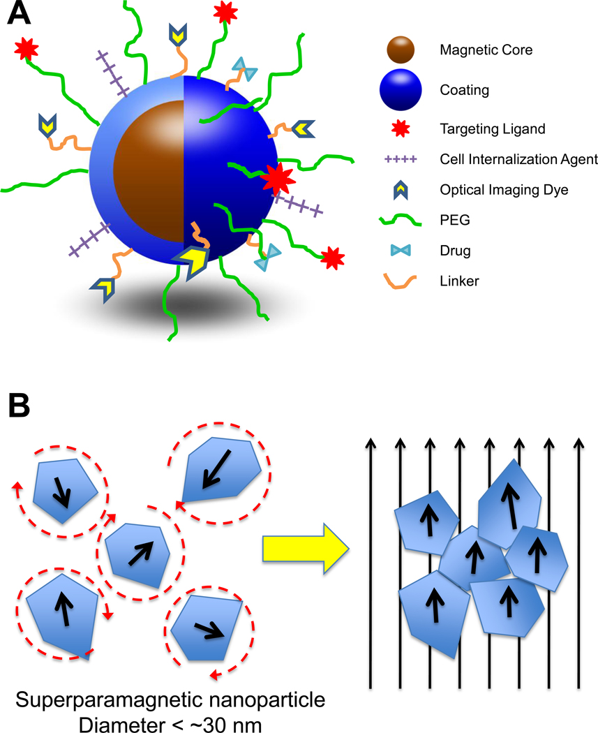
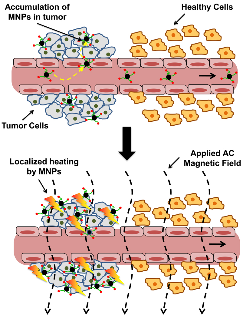
Interest in utilizing magnetic nanoparticles (MNP) for biomedical applications has increased considerably over the past two decades. This excitement has been driven in large part by the success of MNPs as contrast agents in magnetic resonance imaging. The recent investigative trend with respect to cancer has continued down a diagnostic path, but has also turned toward concurrent therapy, giving rise to the distinction of MNPs as potential "theranostics". Here we review both the key technical principles of MNPs and ongoing advancement toward a cancer theranostic MNP. Recent progress in diagnostics, hyperthermia treatments, and drug delivery are all considered. We conclude by identifying current barriers to clinical translation of MNPs and offer considerations for their future development.


Conflict of interest statement

The authors each declare no conflict of interest in this work.

Figures

Figure 1
Figure 1
(A) Schematic representation of core-shell structure of MNPs and multi-functional surface decoration. MNPs consist of an iron oxide core coated with a biocompatible material (e.g. polysaccharide, lipid, proteins, small silane linkers). Functional groups on the surface of coatings are often used to link ligands for molecular targeting, cellular internalization, optical imaging, enhanced plasma residence, and/or therapy. The number of different moieties that decorate the MNP surface impart its multi-functional, “theranostic” character. (B) Illustration of superparamagnetic MNP response to applied magnetic fields. MNPs are comprised of rotating crystals that align with the direction of an applied magnetic field. Crystal reorientation provides for the high magnetic susceptibilities and saturation magnetizations observed with this material. The circular dashed lines around the superparamagnetic nanoparticles on the left illustrate the randomization of their orientation, due to temperature effects, in the absence of a magnetic field.
Figure 2
Figure 2
Reticuloendothelial system (RES) plasma clearance of MNPs. As shown in the conceptual schematic above, opsonin proteins in the circulation adsorb to nanoparticle surfaces, “flagging” them as exogenous materials for plasma clearance. RES tissue macrophages (primarily those of the liver and spleen) recognize flagged MNPs and remove them from the circulation via phagocytosis.
Figure 3
Figure 3
Brain MRI time course of cross-linked starch coated, PEG-modified MNPs in male Fisher 344 rats bearing 9L-glioma brain tumors (12 mg Fe/kg) (Reproduced with permission) [20]. Baseline T2-weighted, fast spin echo images clearly indicate the positioning of the tumor (hyperintense region) in the brain. T2*-weighted, gradient echo images provide qualitative information about MNP presence in the brain/tumor. Sustained negative contrast (hypointensity) in the tumor confirms that D5 (cross-linked starch coated MNP modified with 5000 MW PEG) and D20 (cross-linked starch coated MNP modified with 20000 MW PEG) reside far longer in plasma and provide greater exposure to tumor when compared to D (unmodified, parent starch MNP). Indeed, some hypointensity can still be observed in tumors through 24 h with the PEGylated MNPs.
Figure 4
Figure 4
Conceptual representation of MNP tumor targeting modalities. (A) EPR effect – Unlike that found in normal tissue, tumor vasculature is “leaky” due to fenestrations and gaps between endothelial cells that result from abnormal angiogenesis. MNPs in circulation can passively extravasate through these gaps and enter the tumor interstitium. Poor lymphatic drainage in some tissues helps retain particles in the tumor space. (B) Molecular targeting – ligands (antibodies, peptides, small molecules, etc.) targeted toward moieties overexpressed/uniquely present on the plasma membrane of tumor cells can be used to actively enhance retention of MNPs at the tumor site and can also help to internalize particles into cells via endocytosis. (C) Magnetic targeting – an external magnetic field can be applied to the tumor region, producing a field gradient across the tumor. The magnetic force produced by the gradient actively attracts particles into the tumor space (through comprised vasculature) and helps with subsequent retention. It should be noted that each form of targeting in the figure can be employed simultaneously depending on the particular strategy utilized.
Figure 5
Figure 5
Principle of Magnetic Hyperthermia. Magnetic hyperthermia relies on the preferential accumulation of MNPs at tumors compared to normal tissues. Targeted MNPs delivered to the tumor are exposed to an alternating current (AC) magnetic field, causing nanoparticles to absorb energy by increasing alignment (lower entropy state) with the applied field. This energy is then converted to heat when the particles undergo relaxation. Tumors have been shown to possess thermal hypersensitivity, compared to normal tissues, when heated to temperatures ranging from 41–47°C. The concern for overheating normal tissues remains, however, presenting an important limitation to this method.

References

    1. Walter P, et al. Early Use of PbS Nanotechnology for an Ancient Hair Dyeing Formula. Nano Letters. 2006;6:2215–2219. - PubMed
    1. Reibold M, et al. Materials: Carbon nanotubes in an ancient Damascus sabre. Nature. 2006;444:286–286. - PubMed
    1. Gupta AK, Gupta M. Synthesis and surface engineering of iron oxide nanoparticles for biomedical applications. Biomaterials. 2005;26:3995–4021. - PubMed
    1. Schmid Gn, Wiley I. Nanoparticles: from theory to application. Wiley-VCH ; John Wiley; 2004.
    1. Roca AG, et al. Progress in the preparation of magnetic nanoparticles for applications in biomedicine. Journal of Physics D: Applied Physics. 2009;42:224002.

Publication types

MeSH terms