. 2011 Jun;107(8):1421-6.
doi: 10.1093/aob/mcr074. Epub 2011 Apr 12.

Successive microsporogenesis affects pollen aperture pattern in the tam mutant of Arabidopsis thaliana

Affiliations

Successive microsporogenesis affects pollen aperture pattern in the tam mutant of Arabidopsis thaliana

B Albert et al. Ann Bot. 2011 Jun.

Abstract

Background and aims: The tam (tardy asynchronous meiosis) mutant of Arabidopsis thaliana, which exhibits a modified cytokinesis with a switch from simultaneous to successive cytokinesis, was used to perform a direct test of the implication of cytokinesis in aperture-pattern ontogeny of angiosperm pollen grains. The aperture pattern corresponds to the number and arrangement of apertures (areas of the pollen wall permitting pollen tube germination) on the surface of the pollen grain.

Methods: A comparative analysis of meiosis and aperture distribution was performed in two mutant strains of arabidopsis: quartet and quartet-tam.

Key results: While the number of apertures is not affected in the quartet-tam mutant, the arrangement of the three apertures is modified compared with the quartet, resulting in a different aperture pattern.

Conclusions: These results directly demonstrate the relationship between the type of sporocytic cytokinesis and pollen aperture-pattern ontogeny.


Figures
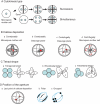
Fig. 1.
Fig. 1.
The four components implicated in aperture-pattern ontogeny. (A) Cytokinesis type. A characteristic dyad stage is observed in successive cytokinesis. Nuclei are represented by small blue circles, callose walls by black bars. (B) Callose deposition. Callose wall formation is illustrated in a tetragonal tetrad (circle with cleavage walls inside). The callose is represented by thick grey lines. Arrows indicate the progression of callose deposition. Red circles represent the location of the last points where callose is deposited between the microspores; the position of these points depends on the way the callose walls are formed. (B-a) Callose deposition starts from the periphery of the pollen mother cell and progresses centripetally toward the centre of microsporocyte. (B-b) Callose deposition starts from the periphery of each cleavage plane and progresses centripetally towards the centre of the cleavage plane. B-c) Callose deposition starts from the centre of each cleavage plane and progresses centrifugally. (B-d) Callose deposition starts from the centre of the microsporocyte and progresses centrifugally. (C) The shape of the tetrad. Blue circles represent pollen grains; the cleavages walls are represented in black and numbered. (C-a) Tetragonal tetrad displays three (in successive cytokinesis) or four cleavage walls (in simultaneous cytokinesis). (C-b) Rhomboidal tetrad displays five cleavage walls. (C-c) Tetrahedral tetrad displays six cleavage walls. (D) Example of aperture distribution within tetrad. (D-a) Last points of callose deposition (red points) within a tetragonal tetrad. In this example the callose deposition corresponds to a centrifugal callose deposition within cleavage planes. (D-b) Polar aperture distribution. The pollen grains are monosulcate with the sulcus oriented toward the red points. (D-c) Grouped aperture distribution. The pollen grains are diporate and the apertures are directly located where the red points are.
Fig. 2.
Fig. 2.
(A–C) Pollen tetrad in quartet strain. Pollen grains are three aperturate – each of the three apertures of a pollen grain is associated with an aperture of another pollen grain. Apertures of a pollen grain are in contact with apertures of the three other pollen grains. Tetrads are tetrahedral. (C-b) Scheme of the tetrad shown in (C-a); big circles represent the microspores, triangles represent apertures. (D–F) Pollen tetrad in quartet-tam strain. Three aperturate pollen grains are associated two by two within the tetrad. All the three apertures of a pollen grain are associated with the three apertures of only one another pollen grain. (D) Tetragonal tetrad. (E) Tetragonal tetrad showing a pollen grain with an abnormal aperture (the upper right). (F-a, -b, -c) Different focal planes on a decussate tetrad; arrows show apertures. (F-d) Scheme of the tetrad shown in (F-a, -b, -c), big circles represent the microspores; black bars represent apertures that are seen by looking from the view of the tetrad in (F-a); grey ones those which are hidden. (A), (D) and (E) are scanning electron micrographs; (B), (C) and (F) are stained with aniline blue. Scale bars = 10 µm.
Fig. 3.
Fig. 3.
(A–C) Microsporogenesis in quartet strain. (A) Centripetal cell plate formation within a cleavage plane; callose deposition started from the border of the tetrad and in the centre (arrows). (B) Two focal planes of a tetrad at the end of cytokinesis; the three last contact points between microspores are indicated by arrows. (C) Tetrahedral tetrad after the completion of cell plate formation. (D–I) Microsporogenesis in the quartet-tam strain. (D) Centripetal cell plate formation within a cleavage plane after the first nuclear division. (E) Dyad. (F, G) Centripetal cell plate formation after the second nuclear division in a decussate tetrad (F) and in a tetragonal tetrad (G). (H-a,b) Two focal planes of a tetrad at the end of cytokinesis showing the three last contact points between two microspores (arrows); it is not possible to see the other two microspores (on left) very well. (I) Tetragonal tetrad after the completion of cell plate formation. All are stained with aniline blue. Scale bars = 10 μm.

References

    1. Albert B, Nadot S, Dreyer L, Ressayre A. The influence of tetrad shape and intersporal callose wall formation on pollen aperture pattern ontogeny in two eudicot species. Annals of Botany. 2010;106:557–564. - PMC - PubMed
    1. Arens K. Prova de calose por meio da microscopia a luz fluorescente e aplicações do metodo. Lilloa. 1949;18:71–75.
    1. Banks H, Stafford P, Crane P. Aperture variation in the pollen of Nelumbo (Nelumbonaceae) Grana. 2007;46:157–163.
    1. Blackmore S, Barnes SH. Garside's rule and the microspore tetrads of Grevillea rosmarinifolia A. Cunningham and Dryandra polycephala Bentham (Proteaceae) Review of Palaeobotany and Palynology. 1995;85:111–121.
    1. Blackmore S, Crane P. The systematic implications of pollen and spore ontogeny. In: Humpries CJ, editor. Ontogeny and systematics. New York, NY: Columbia University Press; 1988. pp. 83–115.