. 2011 Jul 8;286(27):24300-11.
doi: 10.1074/jbc.M111.234104. Epub 2011 Apr 13.

Recognition of multivalent histone states associated with heterochromatin by UHRF1 protein

Affiliations

Recognition of multivalent histone states associated with heterochromatin by UHRF1 protein

Nataliya Nady et al. J Biol Chem. .

Abstract

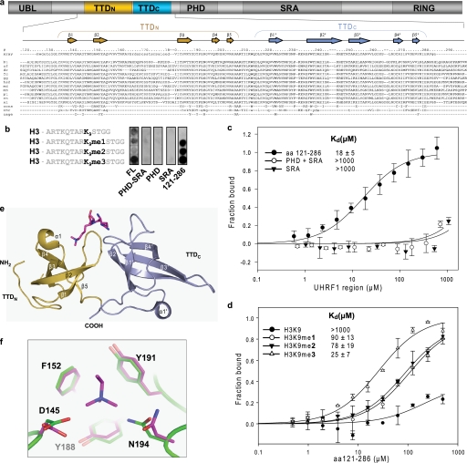
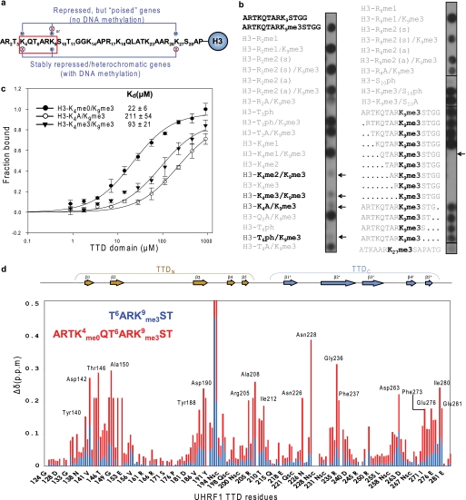
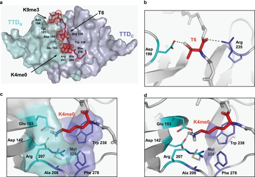
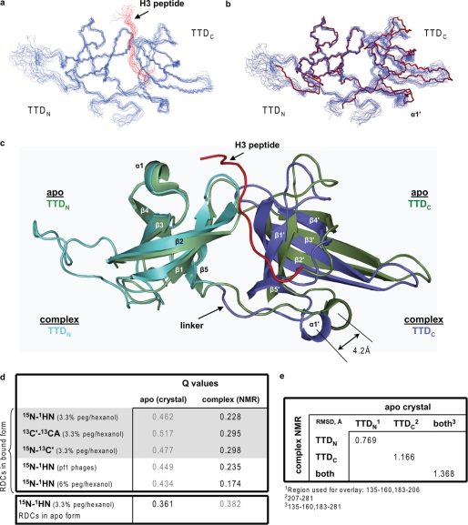
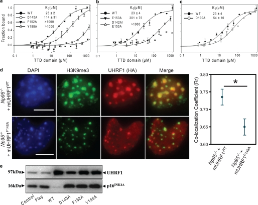
Histone modifications and DNA methylation represent two layers of heritable epigenetic information that regulate eukaryotic chromatin structure and gene activity. UHRF1 is a unique factor that bridges these two layers; it is required for maintenance DNA methylation at hemimethylated CpG sites, which are specifically recognized through its SRA domain and also interacts with histone H3 trimethylated on lysine 9 (H3K9me3) in an unspecified manner. Here we show that UHRF1 contains a tandem Tudor domain (TTD) that recognizes H3 tail peptides with the heterochromatin-associated modification state of trimethylated lysine 9 and unmodified lysine 4 (H3K4me0/K9me3). Solution NMR and crystallographic data reveal the TTD simultaneously recognizes H3K9me3 through a conserved aromatic cage in the first Tudor subdomain and unmodified H3K4 within a groove between the tandem subdomains. The subdomains undergo a conformational adjustment upon peptide binding, distinct from previously reported mechanisms for dual histone mark recognition. Mutant UHRF1 protein deficient for H3K4me0/K9me3 binding shows altered localization to heterochromatic chromocenters and fails to reduce expression of a target gene, p16(INK4A), when overexpressed. Our results demonstrate a novel recognition mechanism for the combinatorial readout of histone modification states associated with gene silencing and add to the growing evidence for coordination of, and cross-talk between, the modification states of H3K4 and H3K9 in regulation of gene expression.


Figures

FIGURE 1.
FIGURE 1.
A novel tandem tudor domain (TTD) within UHRF1 preferentially binds H3 histone tail trimethylated at Lys-9. a, domain composition of UHRF1 includes ubiquitin-like (UBL), TTD, comprising N- and C-terminal subdomains (TTDN, TTDC), PHD, SRA, and RING domains. The human UHRF1 protein sequence (amino acids 119–298) is aligned with that of other species (bt, Bos taurus; cf, Canis familiaris; dr, Danio rerio; fc, Felis catus; gg, Gallus gallus; hs2, Homo sapiens; md, Monodelphis domestica; mm, Mus musculus; pt, Pan troglodytes; rn, Rattus norvegicus; xl, Xenopus laevis); the lowercase sequences are flexible regions in the liganded x-ray structure. The residue numbering corresponds to the human UHRF1 sequence. Drawn above the number line are secondary structure elements and their labels. Residues that are fully conserved (cons) undergo medium and strong (uppercase) and weak (lowercase) changes in chemical shifts with addition of K4me0/K9me3-containing H3 peptide (NMR) and those that form the K9me3 aromatic cage and K4me0 cage (cage) are shown below. b, SPOT-blot peptides corresponding to the amino-terminal tail of histone H3 with or without modification at the Lys-9 position (1, monomethylation; 2, dimethylation; 3, trimethylation) were probed for binding with different UHRF1 domains. The full-length recombinant UHRF1 and a domain spanning residues 121–286 showed clear binding to the histone tail when Lys-9 was methylated. c, FP assays confirm binding of the amino acids 121–286 to a modified peptide corresponding to residues 1–11 of H3 (ARTKQTARKme3ST). d, binding of the UHRF1 construct spanning amino acids 121–286 to the series of peptides that have different methylation state of Lys-9 was measured using FP. Peptides corresponding to residues 1–11 of H3, ARTKQTAR[Kme0/1/2/3]ST with Lys-9 unmodified, mono-, di-, or tri-methylated were used. e, the TTD is shown in ribbon format with TTDN in light brown and TTDC in light blue. A stick representation of the H3K9me3 peptide is shown in magenta; electron density was only observed for three residues, R8K9me3S10. f, close-up view of the aromatic cage in the crystal structures of TTD in its apo form (green) and in complex with H3K9me3 (magenta). Asp-145 and Asn-194 appear to have some plasticity because their side chains rotate to present the apolar faces toward the ligand, consistent with the ability of the domain to interact with lower states of Lys-9 methylation.
FIGURE 2.
FIGURE 2.
Recognition of multivalent histone signatures associated with heterochromatin by UHRF1. a, schematic representation of the multivalent nature of the histone H3 tail. Residues known to be modified in vivo are numbered. Stably repressed genes and heterochromatic regions are characterized by the K4me0/K9me3 state of H3. By contrast, “poised” genes are marked by the bivalent K4me3/K27me3 state accompanied in some cases by K9me3 (72). The region boxed in red is the key sequence studied here. b, a series of H3 peptides were analyzed for binding by the TTD using SPOT-blot arrays. Unmodified H3 peptide (amino acids 1–13) showed no binding, whereas the trimethylated H3K9me3 peptide showed strong binding. The effect of dual modifications, alanine replacement, and deletion (periods) in the background of K9me3 was tested. Arrows indicate peptides for which binding was lost. c, FP assays measuring the binding of the purified TTD to H3 peptides that have Lys-9 trimethylated and Lys-4 that is either mutated or methylated to different degrees. d, composite chemical shift changes versus residue number for UHRF1 TTD domain after binding to H3 tail K4me0/K9me3-containing long peptide (amino acids 1–11) is indicated in red, and shifts observed upon addition of the short peptide (amino acids 6–11) are indicated in blue. Prolines and residues that could not be assigned were given a value of zero. Residues that had a very large chemical shift change such that the corresponding peak in the apo form could not be identified were given a constant value of 0.21 ppm. Residues that were strongly affected are listed on the histogram. A large number of residues (37%) is involved in the interaction with the peptide corresponding to the N-terminal tail of the H3 with trimethylated Lys-9 and Lys-4 unmodified.
FIGURE 3.
FIGURE 3.
Recognition of multivalent sites at the interface between the two Tudor subdomains. a, surface representation of the lowest energy complex NMR structure of TTD bound to the histone H3 tail. The N- and C-terminal Tudor subdomains of UHRF1 are shown in cyan and slate colors, respectively. Key residues on the peptide are shown as red dots. b, the TTD-H3 binding is stabilized by the interaction between the hydroxyl and backbone carbonyl groups of H3 Thr-6 that hydrogen bond with UHRF1 Asp-190 carboxylate and the Arg-235 guanidinium groups, respectively. Only protons participating in hydrogen bonding are shown. c, the H3K4me0 pocket is formed by a hydrophilic wall (residues from TTDN, Asp-142 and Glu-153), an aromatic wall (TTDC, Trp-238 and Phe-278), as well as Met-224 and residues from the linker between the two subdomains Arg-207 and Ala-208. d, detailed interactions between the TTD and K4me0 showing the side chain of Lys-4 is “caged” by two hydrogen bonds.
FIGURE 4.
FIGURE 4.
Structural readjustment of TTDC to accommodate the histone tail. a, solution NMR ensemble (15 structures) of the complex structure showing the polypeptide backbone of TTD (blue) and histone peptide (red). The structures were overlaid over the region within TTD residues 139–161 and 182–279. b, overlay of the solution TTD ensemble (residues 139–161 and 182–206) in the bound form (blue) and x-ray apo structure (red). c, overlay of the apo crystal structure (green) and complex NMR structure (blue). The N and C termini are removed for clarity to better show the linker between the TTD subdomains. The extent of the TTDC shift relative to TTDN upon recognition of the H3K4me0/K9me3 peptide is indicated. d, goodness of the fit between the experimental RDC values measured for either the H3K4me0/K9me3-bound TTD or apo-TTD in solution and the predicted values for the NMR solution structure and apo crystal structures. For couplings highlighted in gray, the same alignment tensor was used for fitting. e, the r.m.s.d. (Å) between the apo crystal structure and lowest energy structure of the ensemble of TTD complex solution structures was calculated with MolMol.
FIGURE 5.
FIGURE 5.
Mutational analysis confirms localization of TTD to heterochromatin in mouse ES cells. a–c, binding of wild-type and mutant TTD forms that disrupt the H3K9me3 binding cage (a), H3K4me0 binding cage (b), or Thr-6 recognition within the recombinant human TTD (c). The fluorescence polarization binding assays were performed using a long, H3K4me0/K9me3-containing histone peptide (amino acids 1–11). d, stably integrated mouse Np95−/− ES cell lines expressing HA-tagged wild-type mUHRF1 (top panel) or mUHRF1F148A (lower panel) were stained for HA, H3K9me3, and DAPI. The F148A mutation in mouse protein is equivalent to F152A in human protein. The extent of mUHRF1 co-localization with H3K9me3 was quantitatively evaluated using 60 independent nuclei selected in an unbiased manner by the ImageJ Nucleus Counter plug-in. The HA-mUHRF1F148A protein shows a significantly reduced correlation coefficient compared with the wild-type protein (p value = 0.0102, average and S.D. are shown). e, effect of UHRF1 wild-type and mutant TTD proteins on p16INK4A protein levels in immortalized human vascular smooth muscle cells (HVTs-SM1). Membranes were probed with an anti-UHRF1 monoclonal antibody, accounting for the presence of an endogenous UHRF1 band in the negative control and vector only transfected cells (FLAG). The blot shown here is representative of at least three independent experiments.

Similar articles

Cited by

References

    1. Denslow S. A., Wade P. A. (2007) Oncogene 26, 5433–5438 - PubMed
    1. Klose R. J., Bird A. P. (2006) Trends Biochem. Sci. 31, 89–97 - PubMed
    1. Bartke T., Vermeulen M., Xhemalce B., Robson S. C., Mann M., Kouzarides T. (2010) Cell 143, 470–484 - PMC - PubMed
    1. Meissner A., Mikkelsen T. S., Gu H., Wernig M., Hanna J., Sivachenko A., Zhang X., Bernstein B. E., Nusbaum C., Jaffe D. B., Gnirke A., Jaenisch R., Lander E. S. (2008) Nature 454, 766–770 - PMC - PubMed
    1. Ooi S. K., Qiu C., Bernstein E., Li K., Jia D., Yang Z., Erdjument-Bromage H., Tempst P., Lin S. P., Allis C. D., Cheng X., Bestor T. H. (2007) Nature 448, 714–717 - PMC - PubMed

Publication types

MeSH terms