. 2011 Jun 17;286(24):21062-72.
doi: 10.1074/jbc.M110.211409. Epub 2011 Apr 13.

Met receptors induce Sam68-dependent cell migration by activation of alternate extracellular signal-regulated kinase family members

Affiliations

Met receptors induce Sam68-dependent cell migration by activation of alternate extracellular signal-regulated kinase family members

Alessia Locatelli et al. J Biol Chem. .

Abstract

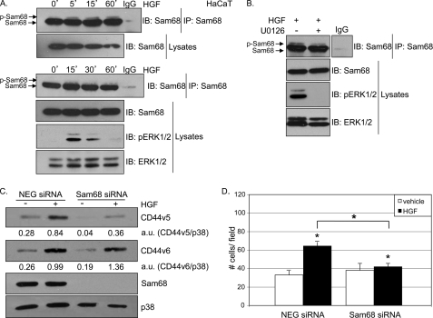
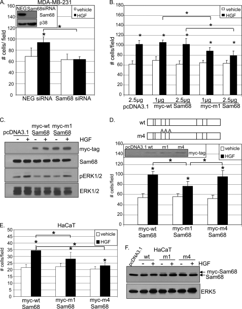
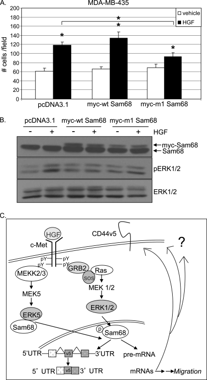
The hepatocyte growth factor (HGF)/Met receptor signaling pathway is deregulated in diverse human malignancies and plays a central role in oncogenesis, tumor progression, and invasive cancer growth. Similarly, altered expression and splicing (i.e. inclusion of variant exon 5, "v5") of the cell adhesion marker, CD44, is associated with advanced cancer phenotypes. We sought to further understand how HGF regulates CD44v5 expression. Immortalized nontumorigenic keratinocyte (HaCaT) cells abundantly express both Met receptors and CD44v5 transmembrane glycoproteins. HGF stimulated CD44v5 protein expression and HaCaT cell migration; these events required activation of the ERK1/2 MAPK module and Sam68, a protein involved in RNA processing, splicing, and v5 inclusion. Similar to HaCaT cells, highly migratory MDA-MB-231 breast cancer cells also required Sam68 expression for HGF-induced migration. However, MDA-MB-231 cell migration occurred independently of ERK1/2 and CD44v5 expression and instead required ERK5 signaling to Sam68. Phospho-mutant, but not WT-Sam68, blocked HGF-induced cell migration in both cell types; MDA-MB-435 cells behaved similarly. These results suggest that Sam68 acts as a convergence point for ERK signaling to cell migration; blockade of phospho-Sam68 may provide a new avenue for therapeutic inhibition of metastatic cancers.


Figures

FIGURE 1.
FIGURE 1.
ERK1/2 activation is required for HGF-induced CD44v5 up-regulation and migration in keratinocyte cells. A, HaCaT cells were serum-starved for 18 h and pretreated with 10 μm of the MEK1/2 inhibitor U0126 for 30 min. Cells were then treated with either vehicle or 50 ng/ml HGF for 4 h. CD44v5 expression and ERK1/2 activation were determined with specific antibodies. Total ERK1/2 served as a loading control. B, Boyden chamber migration assays performed with HGF-stimulated HaCaT cells in the presence or absence of 10 μm U0126. MEK inhibitor was added to both upper and lower chambers (wells). C, Boyden chamber migration assays with HaCaT cells transiently transfected with control or CD44v5 siRNA. Inset, Western blots showing CD44v5 and ERK1/2 expression upon CD44v5 knockdown. Forty eight hours post-transfection, cells were harvested and assayed for HGF-induced migration. * denotes significance (p < 0.05) relative to vehicle between indicated groups, determined by unpaired Student's t test. § denotes no statistical difference relative to vehicle.
FIGURE 2.
FIGURE 2.
Sam68 is required for HGF-induced MAPK-dependent CD44v5 up-regulation and keratinocyte cell migration. A, HaCaT cells were starved for 18 h and treated with either vehicle or 50 ng/ml HGF for 5, 15, and 60 min (top panel) or 15, 30, and 60 min (bottom panel). B, HaCaT cells were starved for 18 h, pretreated with either vehicle or 10 μm of the MEK inhibitor U0126 for 30 min, and treated with 50 ng/ml HGF for 15 min. Cellular lysates (A and B) were immunoprecipitated (IP) for Sam68 and immunoblotted (IB) using Sam68-specific antibodies. Normal rabbit IgG was used as specificity control. ERK1/2 activation was evaluated using phospho-specific antibodies; total ERK1/2 served as a loading control. C, cells transiently expressing either control or Sam68 siRNA were starved for 18 h, induced with 50 ng/ml HGF for 4 h, and Western-blotted (IB) with antibodies specific to CD44v5, CD44v6, and Sam68. Total p38 served as a loading control. Densitometry analysis was performed and indicated below the proper lane as a ratio between CD44v5 and p38 intensity or CD44v6 and p38. D, control and Sam68 siRNA-expressing cells were assayed for their ability to migrate in the presence of HGF. * denotes significance (p < 0.05) relative to vehicle and between indicated groups, as determined by unpaired Student's t test.
FIGURE 3.
FIGURE 3.
HGF-induced migration requires Sam68 phosphorylation. A, top panel, schematic representation of MAPK consensus (PXX(S/T)P) sites on Myc-tagged WT-Sam68 and phospho-mutant m1 wherein all eight Thr or Ser residues are replaced by Ala (30). Bottom panel, cells were transiently transfected with 2.5 μg of vector and 1 and 2.5 μg of Myc-tagged wild-type Sam68 or Myc-tagged phospho-mutant m1. After 48 h, cells were assayed for HGF-induced migration in Boyden chambers. * denotes significance (p < 0.05) relative to vehicle and between indicated groups, as determined by unpaired Student's t test. B, Western blots showing Myc-tagged Sam68 and endogenous Sam68 expression; C, HGF-induced activation of ERK1/2. Total ERK1/2 served as a loading control.
FIGURE 4.
FIGURE 4.
MAPK signaling mediates Sam68 phosphorylation and migration in MDA-MB-231 breast cancer cells. A, HaCaT and MDA-MB-231 cells were starved for 18 h and treated with either vehicle or 50 ng/ml HGF for 4 h. CD44v5 expression was determined with specific antibodies. Total p38 served as a loading control. B, MDA-MB-231 cells were starved for 18 h, pretreated with either vehicle or 10 μm MEK1/2 inhibitor (U0126) for 30 min, and subsequently treated with 50 ng/ml HGF for 30 min. Sam68 was immunoprecipitated (IP) from whole cell lysates and then subjected to Western blotting (IB) using Sam68-specific antibodies. Normal rabbit IgG served as a specificity control. Upshifted Sam68 band indicates multisite phosphorylation. C, Boyden chamber migration assays preformed with MDA-MB-231 stimulated with HGF in the presence or absence of MEK1/2 inhibitor, U0126. Inset shows HGF-induced ERK1/2 activation; ERK1/2 protein levels served as loading control. * denotes significance (p < 0.05) relative to vehicle and between indicated groups, as determined by unpaired Student's t test.
FIGURE 5.
FIGURE 5.
ERK5 mediates breast cancer cell migration through ERK5-Sam68 complex dissociation. MDA-MB-231 cells were pretreated with either 10 μm U0126 for 30 min (A) or 10 nm or 10 μm PD (B) and then treated with either vehicle or 50 ng/ml HGF for 15 min. ERK1/2 and ERK5 activation was evaluated using phospho-specific and total specific antibodies, respectively; total ERK1/2 served as a loading control. C, MDA-MB-231 cells were assayed for migration in response to HGF in the presence or absence of either low (10 nm) or high (10 μm) concentrations of PD. D, Boyden migration assays were performed using cells transiently expressing control or ERK5 siRNA (inset shows ERK5 and a nonspecific protein loading control) and stimulated with HGF. * denotes significance (p < 0.05) relative to vehicle and between indicated groups, as determined by unpaired Student's t test. § denotes no statistical difference relative to vehicle. E, cells were treated with either vehicle or 50 ng/ml HGF for 15–30 min. ERK5 was immunoprecipitated (IP) from whole cell lysates using total ERK5-specific antibodies. ERK5 immunoprecipitates were then subjected to Western blotting (IB) with total ERK5 and Sam68-specific antibodies. F, cells were pretreated for 30 min with low (10 nm) or high (10 μm) concentration of PD, followed by 15 min of HGF treatment. ERK5 was immunoprecipitated from whole cell lysates using total ERK5-specific antibodies and Western-blotted with ERK5 and Sam68-specific antibodies. ERK1/2 and ERK5 activation was evaluated using phospho-specific and total specific antibodies, respectively.
FIGURE 6.
FIGURE 6.
Sam68 mediates HGF-induced migration through MAPK-directed phosphorylation sites in breast cancer cells. A, MDA-MB-231 cells expressing either control or Sam68 siRNA were assayed for HGF-induced migration. Inset, Western blots showing Sam68 knockdown and p38 loading control. B, MDA-MB-231 breast cancer cells were transiently transfected with 2.5 μg of vector or 1–2.5 μg of Myc-tagged WT- or m1-Sam68 and assayed in Boyden chambers for HGF induced migration. C, expression of Myc-tagged and endogenous Sam68 protein and ERK1/2 activation were detected using specific antibodies; total ERK1/2 served as a loading control. D, MDA-MB-231 cells transiently transfected with 2.5 μg of Myc-tagged WT-, m1-, and m4-Sam68 were subjected to HGF-induced Boyden chamber migration assays. Schematic representations of Myc-tagged WT- and m4-Sam68 are shown. Inset, transfected Sam68 protein levels were detected using Myc tag-specific antibodies. E, HaCaT cells were transiently transfected as in D and assayed for migration. * denotes significance (p < 0.05) relative to vehicle and between indicated groups, as determined by unpaired Student's t test. F, endogenous and Myc-tagged WT- and m4-Sam68 and total ERK5 were visualized by Western blotting using specific antibodies. Slower running (higher migrating) Sam68 bands indicate Myc-tagged proteins.
FIGURE 7.
FIGURE 7.
Phospho-Sam68 mediates HGF-induced Met+ cancer cell migration. A, MDA-MB-435 cells were transiently transfected as described above with vector, WT-, or m1-Sam68 and assayed for HGF-induced migration. B, Western blot showing transfected and endogenous Sam68 protein expression and MAPK activation. Endogenous and Myc-tagged WT- and m4-Sam68 were visualized by Western blotting using Sam68-specific antibodies. Slower running bands indicate Myc-tagged proteins. ERK1/2 activation was evaluated using phospho-specific antibodies; total ERK1/2 served as a loading control. * denotes significance (p < 0.05) relative to vehicle between indicated groups, as determined by unpaired Student's t test. C, mechanism of action of Sam68 downstream of activated Met receptors. Sam68 is the common effector of HGF-induced migration in keratinocyte and breast cancer cells. Contrary to ERK1/2- and CD44v5-dependent keratinocyte migration, breast cancer cell migration occurs independently of CD44v5 expression via an ERK1/2- or ERK5-dependent mechanism, suggesting a role for phospho-Sam68 in the regulation of splicing of multiple gene targets important for HGF-induced cell migration.

Similar articles

Cited by

References

    1. Nakamura T., Nawa K., Ichihara A. (1984) Biochem. Biophys. Res. Commun. 122, 1450–1459 - PubMed
    1. Russell W. E., McGowan J. A., Bucher N. L. (1984) J. Cell. Physiol. 119, 183–192 - PubMed
    1. Stoker M., Gherardi E., Perryman M., Gray J. (1987) Nature 327, 239–242 - PubMed
    1. Jiang W., Hiscox S., Matsumoto K., Nakamura T. (1999) Crit. Rev. Oncol. Hematol. 29, 209–248 - PubMed
    1. Birchmeier C., Birchmeier W., Gherardi E., Vande Woude G. F. (2003) Nat. Rev. Mol. Cell Biol. 4, 915–925 - PubMed

Publication types

MeSH terms