. 2011 Jun;85(12):5764-72.
doi: 10.1128/JVI.00342-11. Epub 2011 Apr 13.

Significant protection against high-dose simian immunodeficiency virus challenge conferred by a new prime-boost vaccine regimen

Affiliations

Significant protection against high-dose simian immunodeficiency virus challenge conferred by a new prime-boost vaccine regimen

John B Schell et al. J Virol. 2011 Jun.

Abstract

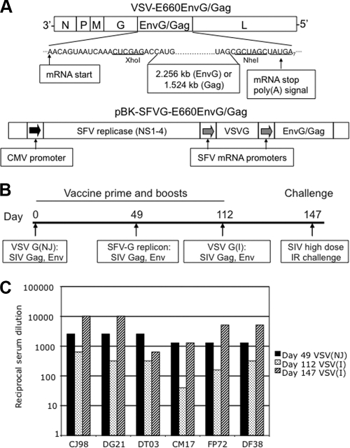
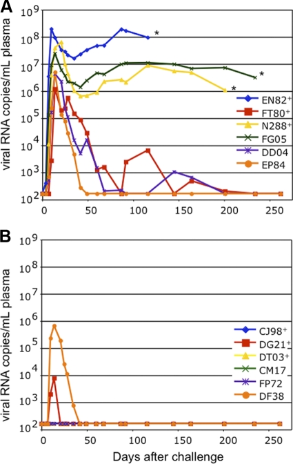
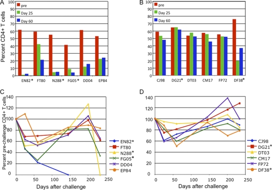
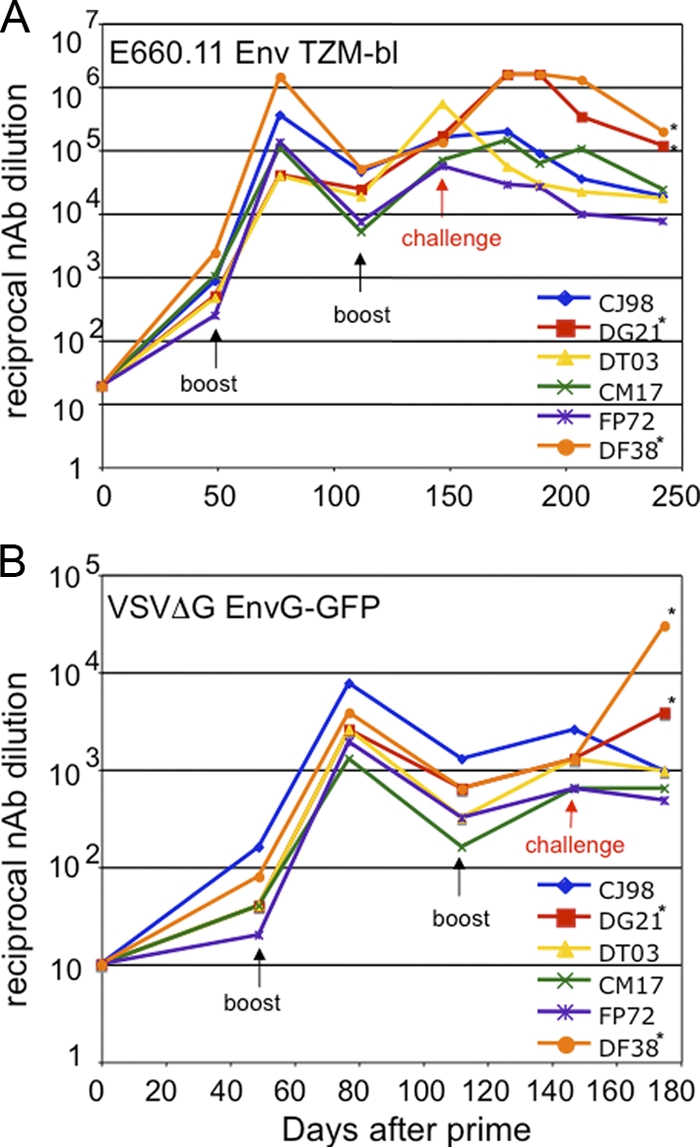
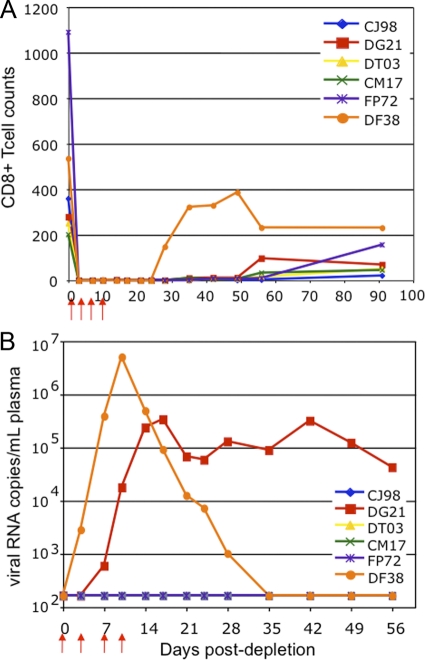
We constructed vaccine vectors based on live recombinant vesicular stomatitis virus (VSV) and a Semliki Forest virus (SFV) replicon (SFVG) that propagates through expression of the VSV glycoprotein (G). These vectors expressing simian immunodeficiency virus (SIV) Gag and Env proteins were used to vaccinate rhesus macaques with a new heterologous prime-boost regimen designed to optimize induction of antibody. Six vaccinated animals and six controls were then given a high-dose mucosal challenge with the diverse SIVsmE660 quasispecies. All control animals became infected and had peak viral RNA loads of 10(6) to 10(8) copies/ml. In contrast, four of the vaccinees showed significant (P = 0.03) apparent sterilizing immunity and no detectable viral loads. Subsequent CD8(+) T cell depletion confirmed the absence of SIV infection in these animals. The two other vaccinees had peak viral loads of 7 × 10(5) and 8 × 10(3) copies/ml, levels below those of all of the controls, and showed undetectable virus loads by day 42 postchallenge. The vaccine regimen induced high-titer prechallenge serum neutralizing antibodies (nAbs) to some cloned SIVsmE660 Env proteins, but antibodies able to neutralize the challenge virus swarm were not detected. The cellular immune responses induced by the vaccine were generally weak and did not correlate with protection. Although the immune correlates of protection are not yet clear, the heterologous VSV/SFVG prime-boost is clearly a potent vaccine regimen for inducing virus nAbs and protection against a heterogeneous viral swarm.


Figures

Fig. 1.
Fig. 1.
VSV and SFVG vaccine constructs, vaccination schedule, and immune responses to the vectors. (A) Diagram of the VSV and SFVG vector constructs. The VSV gene insertion sites are indicated by the flanking transcription and translation start and stop sequences. Positions of the same gene inserts are shown in the SFVG vector relative to the two subgenomic mRNA promoters. (B) Schedule for vaccination and SIVsmE660 challenge. Days are given as days postprime. IR, intrarectal. (C) Postprime neutralizing antibody titers to the two VSV G proteins present in the vectors. Titers represent the reciprocal of the serum dilution that completely neutralized 100 PFU of the indicated VSV Indiana or New Jersey serotype viruses.
Fig. 2.
Fig. 2.
Vaccine group animals appear to be uninfected or have low viral loads compared to those of control animals. The graphs show the number of plasma viral RNA copies per ml of sera in control group animals (A) and vaccine group animals (B) on the indicated days following high-dose mucosal (rectal) challenge with the SIVsmE660 swarm. Control animals marked with an asterisk were euthanized when they developed AIDS. The plus symbols indicate the three animals in each group that carried the Mamu-A*01 allele. No viral loads were detectable in 4 of the 6 vaccine group animals at any time following challenge. The difference in peak viral loads between the two groups was significant (P < 0.001; two-tailed Mann-Whitney test).
Fig. 3.
Fig. 3.
Gut and peripheral blood CD4+ T cell levels in control and vaccine group animals. Bar graphs for the control group (A) and vaccine group (B) show the percentages of CD3+ gut lymphocytes that are CD4+ at the day of challenge and at 25 and 60 days postchallenge. Line graphs for the control group (C) and vaccine group (D) show the CD4+ T cell counts in the blood, expressed as percentages of the average prechallenge CD4+ T cell counts. Asterisks indicate the control animals with the highest virus loads and the two vaccine group animals that were initially infected but controlled their virus loads.
Fig. 4.
Fig. 4.
Pre- and postchallenge serum nAb responses from the vaccine group animals. (A) Serum nAb to a pseudovirus expressing the E660.11 envelope, measured by the TZM-bl assay. (B) nAb responses to the EnvG protein present in the vaccine using a VSVΔG surrogate virus. The initial VSV vector prime was on day 0. The black arrows indicate the days of the SFVG and VSV vector boosts. The days of SIVsmE660 challenge are indicated by the red arrows. Note that the two infected animals in the vaccine group (indicated by asterisks) showed a strong anamnestic response in antibody production following challenge in both assays.
Fig. 5.
Fig. 5.
Tetramer and ELISPOT assays of vaccine group Mamu-A*01+ PBMCs. (A) Results from tetramer staining analysis of CD8+ T cells recognizing the immunodominant Gag p11C epitope. Data are presented as the percentages of CD3+ CD8+ cells that are also p11C tetramer positive. (B) Levels of IFN-γ secreting specific for SIV Gag, as detected by ELISPOT assay. Data are presented as the number of spot-forming cells (SFC) per 1 × 10⁶ cells.
Fig. 6.
Fig. 6.
CD8+ T cell depletion in vaccinees shows virus rebound in only the 2/6 animals that showed initial viral loads. All vaccine group animals were depleted for CD8+ T cells by treatment with rhesus anti-CD8 monoclonal antibody administered on the days indicated by the red arrows. (A) Rapid and sustained CD8+ T cell depletion in all animals after the antibody treatment. Values are represented as the total CD8+ cells counts per μl of blood. (B) Postdepletion analysis of plasma viral RNA levels.

Similar articles

Cited by

References

    1. Barefoot B., et al. 2008. Comparison of multiple vaccine vectors in a single heterologous prime-boost trial. Vaccine 26:6108–6118 - PMC - PubMed
    1. Barouch D. H., et al. 2002. Eventual AIDS vaccine failure in a rhesus monkey by viral escape from cytotoxic T lymphocytes. Nature 415:335–339 - PubMed
    1. Boritz E., Gerlach J., Johnson J. E., Rose J. K. 1999. Replication-competent rhabdoviruses with human immunodeficiency virus type 1 coats and green fluorescent protein: entry by a pH-independent pathway. J. Virol. 73:6937–6945 - PMC - PubMed
    1. Buchbinder S. P., et al. 2008. Efficacy assessment of a cell-mediated immunity HIV-1 vaccine (the Step study): a double-blind, randomised, placebo-controlled, test-of-concept trial. Lancet 372:1881–1893 - PMC - PubMed
    1. Casimiro D. R., et al. 2005. Attenuation of simian immunodeficiency virus SIVmac239 infection by prophylactic immunization with DNA and recombinant adenoviral vaccine vectors expressing Gag. J. Virol. 79:15547–15555 - PMC - PubMed

Publication types

MeSH terms