. 2011 Jun;85(12):5853-63.
doi: 10.1128/JVI.02425-10. Epub 2011 Apr 13.

The replisome pausing factor Timeless is required for episomal maintenance of latent Epstein-Barr virus

Affiliations

The replisome pausing factor Timeless is required for episomal maintenance of latent Epstein-Barr virus

Jayaraju Dheekollu et al. J Virol. 2011 Jun.

Abstract

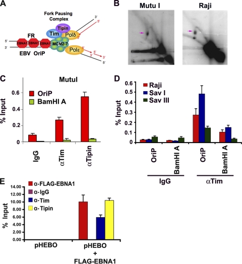
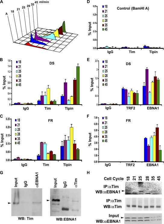
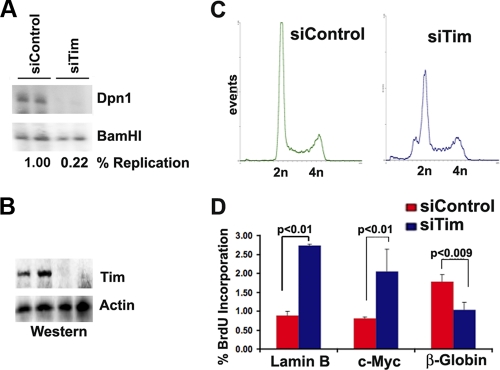
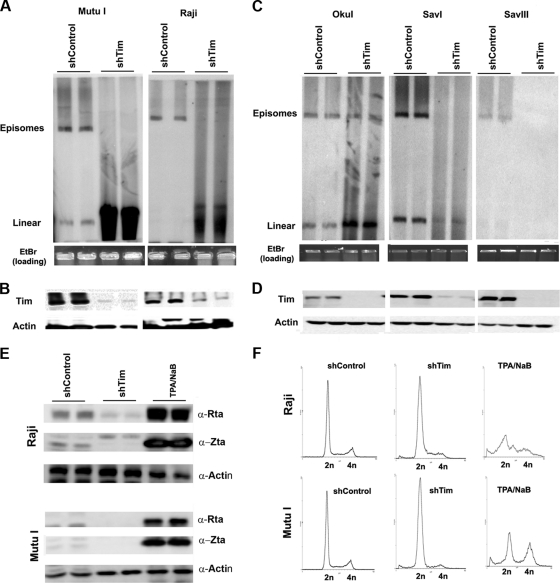
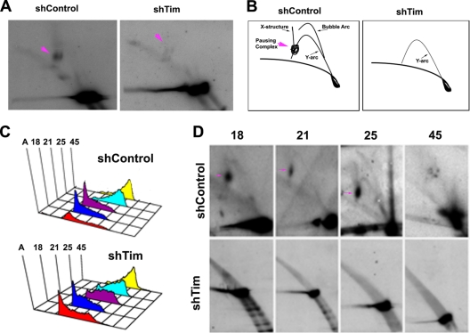
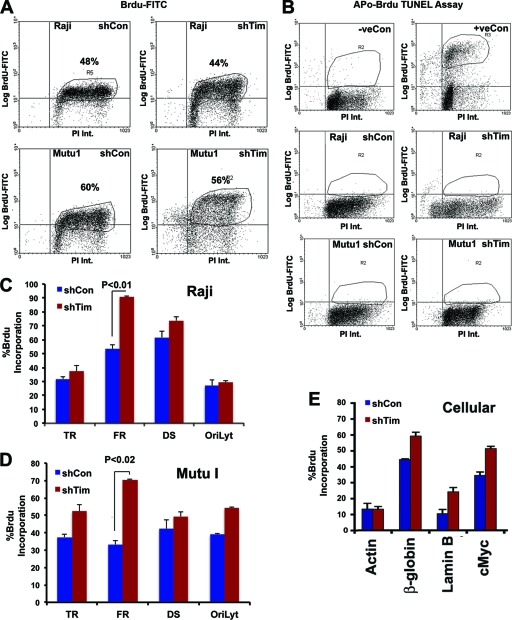
The Epstein-Barr virus (EBV) genome is maintained as an extrachromosomal episome during latent infection of B lymphocytes. Episomal maintenance is conferred by the interaction of the EBV-encoded nuclear antigen 1 (EBNA1) with a tandem array of high-affinity binding sites, referred to as the family of repeats (FR), located within the viral origin of plasmid replication (OriP). How this nucleoprotein array confers episomal maintenance is not completely understood. Previous studies have shown that DNA replication forks pause and terminate with high frequency at OriP. We now show that cellular DNA replication fork pausing and protection factors Timeless (Tim) and Tipin (Timeless-interacting protein) accumulate at OriP during S phase of the cell cycle. Depletion of Tim inhibits OriP-dependent DNA replication and causes a complete loss of the closed-circular form of EBV episomes in latently infected B lymphocytes. Tim depletion also led to the accumulation of double-strand breaks at the OriP region. These findings demonstrate that Tim is essential for sustaining the episomal forms of EBV DNA in latently infected cells and suggest that DNA replication fork protection is integrally linked to the mechanism of plasmid maintenance.


Figures

Fig. 1.
Fig. 1.
Replication fork pausing factors associate with the OriP region of EBV. (A) Schematic model of Tim and Tipin bound to replication fork components at EBNA1 binding sites of OriP. (B) 2D neutral agarose gel of OriP replicating DNA in Mutu I (left panel) or Raji (right panel) cells. Replication fork pause structures are indicated by the pink arrows. (C) ChIP assay of Mutu I cells with antibodies to Tim (αTim), Tipin (αTipin), or control IgG. ChIP DNA was quantified by quantitative PCR (qPCR) at OriP or the control BamHI A region. (D) ChIP assay with anti-Tim or control antibody at either OriP or the control BamHI A DNA region for either Raji (red), Sav I (blue), or Sav III (green) cells. (E) 293 cells were transfected with pHEBO with either pFLAG-EBNA1 or the pFLAG-vector control and then assayed by ChIP at 48 h posttransfection. ChIP was performed with antibodies against FLAG (red), Tim (blue), Tipin (yellow), or control IgG (purple) and assayed for OriP DNA by real-time PCR. Error bars represent standard deviations from the mean for at least three experimental replicates.
Fig. 2.
Fig. 2.
Cell-cycle-dependent association of Tim and Tipin at OriP. (A) Mutu I cells were fractionated by centrifugal elutriation and then assayed by FACS. Elutriation fractions are indicated in ml/min. (B) ChIP assay with anti-Tim, anti-Tipin, or control IgG at the DS region for each stage of the cell cycle as indicated. (C) Same as in panel B, except for analysis at the FR region. (D) Same as in panel B, except for analysis at the control BamHI A region. (E) ChIP assay with anti-EBNA1, anti-TRF2, or control IgG at the OriP DS region for each stage of the cell cycle, as indicated. (F) Same as in panel E, except for analysis at the FR region. (G) Co-IP analysis of EBNA1 and Tim in asynchronous Mutu I cells. EBNA1 or control IgG IPs were assayed by Western blotting (WB) for Tim protein (left panel). Tim or control IgG IPs were assayed by Western blotting for EBNA1 protein (right panel). Arrowheads indicate the expected sizes of the indicated proteins. αEBNA1, anti-EBNA1; αTim, anti-Tim. (H) Cell cycle fractions from Mutu I cells were subject to IP with antibody to Tim and then assayed by Western blotting with anti-EBNA1 (top panel) or anti-Tim (middle panel). The EBNA1 input for each cell cycle fraction is shown in the lower panel.
Fig. 3.
Fig. 3.
Inhibition of OriP DNA replication by Tim depletion. (A) Transient DNA replication assay for OriP-EBNA1 plasmid in 293 cells cotransfected with siControl or siTim. Plasmid DNA was isolated 72 h posttransfection, and subjected to restriction enzyme digestion with DpnI-BamHI (top panel) or BamHI alone (lower panel). Quantification of ratio between DpnI-resistant and total BamHI-cut DNA is used to measure the percentage of DNA replication. (B) Western blot of cell extracts from panel A with antibodies to Tim (top panel) or actin (lower panel). (C) FACS analysis of propidium iodide-stained cells for cell cycle profile analysis of siControl- and siTim-transfected cells used for replication assays shown in panel A. (D) BrdU incorporation studies of 293 cells after transfection with siControl or siTim. BrdU incorporation was analyzed at 48 h posttransfection and analyzed for cellular origins at the lamin B, c-Myc, or β-globin loci.
Fig. 4.
Fig. 4.
Loss of EBV episomal forms in Burkitt's lymphoma cell lines after Tim depletion. (A) Pulsed-field electrophoresis gel analysis of Mutu I (left) or Raji (right) cells after infection with shControl or shTim lentivirus. EBV DNA was detected by Southern blotting. Episomes and linear forms of EBV genomes are indicated. EtBr, ethidium bromide. (B) Western blot of cells used for pulsed-field analysis shown in panel A. Antibodies for Tim (top panel) or actin (lower panel) are indicated. (C) Same analysis as described in panel A for Oku I, Sav I, and Sav III cells. (D) Western blots showing Tim (top) and actin (lower) levels for cells analyzed in panel C. (E) Western blot analysis of EBV lytic proteins Rta, Zta, and control actin for Raji and Mutu I cells after infection with shControl or shTim, or after treatment with tetradecanoyl phorbol acetate (TPA) and NaB for 48 h, as indicated. α-, anti-. (F) FACS analysis of cell cycle profiles for Raji and Mutu I cells after infection with shControl or shTim or after treatment with TPA and NaB for 48 h.
Fig. 5.
Fig. 5.
Tim depletion causes a loss of replication fork structures at OriP. (A) 2D neutral agarose gel electrophoresis and Southern blot analysis of OriP DNA in Mutu I cells after infection with shControl (left panel) or shTim (right panel) lentivirus. Pink arrows indicate replication fork pausing structures. (B) Schematic interpretation of 2D neutral agarose gels shown in panel A. (C) FACS analysis of cell cycle fractions from centrifugal elutriation after shControl lentivirus infection of Mutu I cells. (B) Same as in panel A, except for shTim lentivirus infection. (D) 2D neutral agarose gel analysis of OriP DNA from shControl (top row)- or shTim (bottom row)-infected Mutu I cells. Cell cycle fractions 18, 21, 25, and 45, are indicated. Replication fork pausing structures are indicated by pink arrows.
Fig. 6.
Fig. 6.
Accelerated DNA replication through FR in Tim-depleted cells. (A) FACS analysis of Raji (top panels) or Mutu I (lower panels) cells infected with shControl (left panels) or shTim (right panels) after pulse-labeling with BrdU for 30 min, followed by staining with propidium iodide (PI). BrdU intensity (Int.) was monitored by anti-BrdU-conjugated fluorescein isothiocyanate (FITC) (y axis), and PI was monitored on the x axis. (B) Apo-BrdU TUNEL assay for Raji (middle) or Mutu I (lower) after shControl (left) or shTim (right) infection. Control samples were camptothecin-treated (+) or untreated (−) HL60 cells provided by the manufacturer (top panels). (C) BrdU-ChIP assay for Raji cells after infection with shControl (blue) or shTim (red). BrdU incorporation was assayed at EBV locations for terminal repeats (TR), FR, DS, and OriLyt, as indicated. (D) Same as in panel C, except Mutu I cells were analyzed. (E) Same as in panel D, except BrdU was assayed at the cellular loci for actin, β-globin, lamin B, and c-Myc in Mutu I cells. Error bars indicate standard deviations from the mean, and P values were determined by Chi-square test.
Fig. 7.
Fig. 7.
Tim prevents the formation of double-strand breaks at OriP. (A) Mutu I cells (left) or Raji cells (right) were infected with either shControl or shTim, as indicated. Total intact DNA was extracted with agarose plugs digested with BamHI and then assayed by Southern blot analysis with probes for OriP (BamHI C) (A) or OriLyt (detecting OriLyt duplicated sequence in BamHI d and BamHI H) (B). Double-strand breaks are indicated by arrowhead. Molecular mass markers are indicated on the left. A schematic of the BamHI C probe and fragments are shown below panel A. Predicted breakpoints in the OriP region are indicated by the vertical arrow, gel fragments are indicated by horizontal lines, and numbers are derived from the EBV coordinates in GenBank accession no. AJ507799.

Similar articles

Cited by

References

    1. Aiyar A., Aras S., Washington A., Singh G., Luftig R. B. 2009. Epstein-Barr Nuclear Antigen 1 modulates replication of oriP-plasmids by impeding replication and transcription fork migration through the family of repeats. Virol. J. 6:29. - PMC - PubMed
    1. Allers T., Lichten M. 2000. A method for preparing genomic DNA that restrains branch migration of Holliday junctions. Nucleic Acids Res. 28:e6. - PMC - PubMed
    1. Bairwa N. K., Zzaman S., Mohanty B. K., Bastia D. 2010. Replication fork arrest and rDNA silencing are two independent and separable functions of the replication terminator protein Fob1 of Saccharomyces cerevisiae. J. Biol. Chem. 285:12612–12619 - PMC - PubMed
    1. Ballestas M. E., Chatis P. A., Kaye K. M. 1999. Efficient persistence of extrachromosomal KSHV DNA mediated by latency-associated nuclear antigen. Science 284:641–644 - PubMed
    1. Ballestas M. E., Kaye K. M. 2001. Kaposi's sarcoma-associated herpesvirus latency-associated nuclear antigen 1 mediates episome persistence through cis-acting terminal repeat (TR) sequence and specifically binds TR DNA. J. Virol. 75:3250–3258 - PMC - PubMed

Publication types

MeSH terms

LinkOut - more resources