. 2011 Jun 15;22(12):2094-105.
doi: 10.1091/mbc.E11-01-0054. Epub 2011 Apr 13.

AP-1 and clathrin are essential for secretory granule biogenesis in Drosophila

Affiliations

AP-1 and clathrin are essential for secretory granule biogenesis in Drosophila

Jason Burgess et al. Mol Biol Cell. .

Abstract

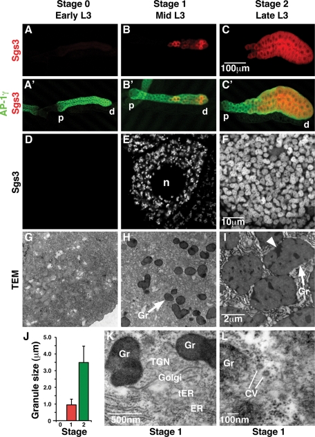
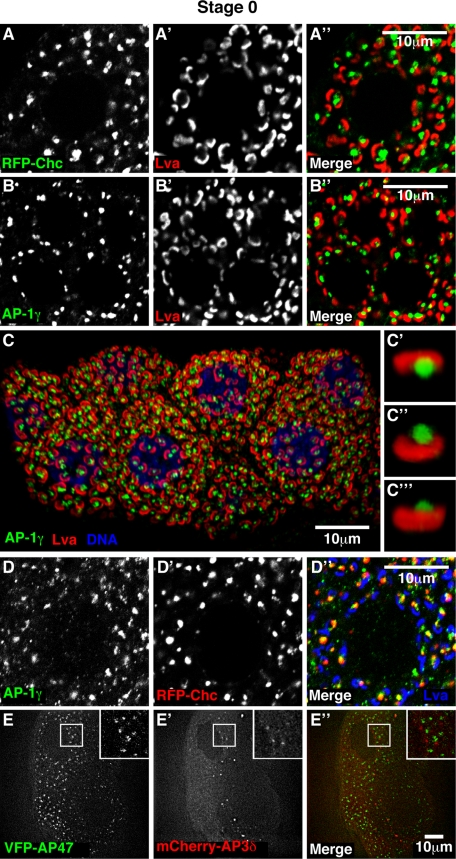
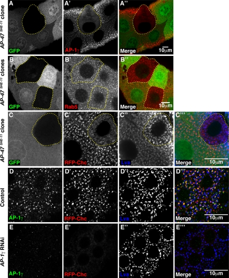
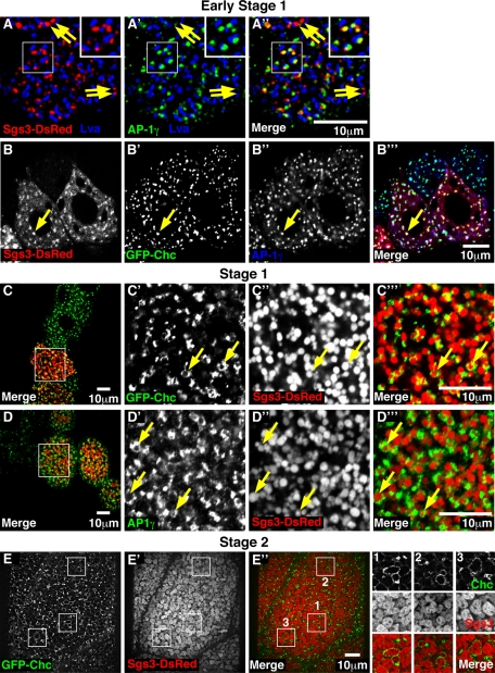
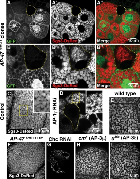
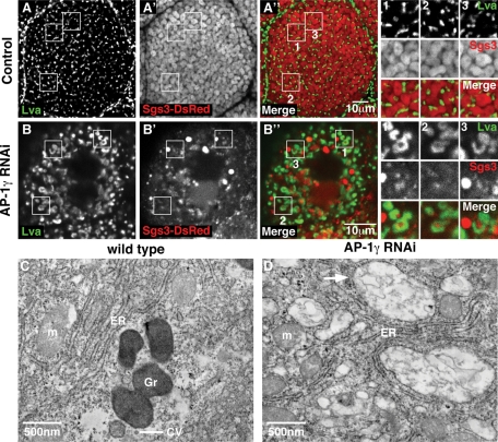
Regulated secretion of hormones, digestive enzymes, and other biologically active molecules requires the formation of secretory granules. Clathrin and the clathrin adaptor protein complex 1 (AP-1) are necessary for maturation of exocrine, endocrine, and neuroendocrine secretory granules. However, the initial steps of secretory granule biogenesis are only minimally understood. Powerful genetic approaches available in the fruit fly Drosophila melanogaster were used to investigate the molecular pathway for biogenesis of the mucin-containing "glue granules" that form within epithelial cells of the third-instar larval salivary gland. Clathrin and AP-1 colocalize at the trans-Golgi network (TGN) and clathrin recruitment requires AP-1. Furthermore, clathrin and AP-1 colocalize with secretory cargo at the TGN and on immature granules. Finally, loss of clathrin or AP-1 leads to a profound block in secretory granule formation. These findings establish a novel role for AP-1- and clathrin-dependent trafficking in the biogenesis of mucin-containing secretory granules.


Figures

FIGURE 1:
FIGURE 1:
Glue granule biogenesis is developmentally regulated. (A–C′) Confocal micrographs of whole third-instar larval (L3) salivary glands expressing Sgs3-DsRed (red) and stained for AP-1γ (green), showing developmental timing of Sgs3-DsRed expression from stage 0 (no granules) through stage 1 (initiation of granule production) to stage 2 (fully mature granules or glands). AP-1γ is expressed in all cells of the salivary gland throughout development, whereas Sgs3-DsRed is first detected in distal (d) mid-L3 salivary gland cells (B, B′) and is expressed in more proximal (p) cells as development proceeds (C, C′). (D–F) Confocal micrographs of individual salivary gland cells showing developmental expression of Sgs3-DsRed. Sgs3-DsRed is not expressed in stage 0 (D). In stage 1, granules surround the nucleus (n) and appear uniformly small (E). In stage 2, granules are larger and occupy most of the cytoplasmic space (F). (G–I) Transmission electron micrographs (TEM) of L3 salivary glands staged using the Sgs3-DsRed marker. No granules were detected in stage 0 (G). Glue granule (Gr) maturation observed by TEM (H, I) parallels that seen by Sgs3-DsRed, validating this marker for following glue granule biogenesis (E, F). (J) Granules increase in size over time, from an average length of 1.0 μm ± 0.3 (n = 91) at stage 1 (red bar) to a maximum length of 3.5 μm ± 1.0 (n = 54) at stage 2 (green bar). (K, L) TEM of stage 1 salivary gland cells. Rough ER, transitional ER (tER), Golgi, and TGN (defined morphologically as in the work of Thomopoulos et al., 1992; Kondylis and Rabouille, 2009) are present near small glue granules (Gr) (K). Coated vesicles (CV) were also observed near glue granules (Gr) (L).
FIGURE 2:
FIGURE 2:
Clathrin heavy chain and the clathrin adaptor AP-1 colocalize at the trans-Golgi network. Confocal micrographs of stage 0 salivary gland cells. (A–A″) RFP-Chc (green) localizes adjacent to, but does not overlap with, the cis-Golgi marker Lva (red). (B–B″) Endogenous AP-1γ (green) localizes adjacent to Lva (red). (C–C″) Projection of a series of spinning-disk confocal images of salivary gland cells stained for AP-1γ (green), Lva (red), and DNA (stained with DAPI; blue) reveals numerous Golgi bodies scattered throughout the cytoplasm (C). A three-dimensional rotation of a single Golgi body shows AP-1γ (green) adjacent to the cup-shaped Lva-positive cis-Golgi (red) (C’–C″). Images were generated from Z stacks of 28 (C) or 5 (C’–C″) optical sections acquired at a distance of 0.3 μm (see Materials and Methods). (D–D″) AP-1γ (green) and RFP-Chc (red) colocalize adjacent to Lva (blue). Colocalization of AP-1γ and RFP-Chc appears yellow in the merged image. (E–E″) Spinning-disk confocal images reveal that VFP-AP-47 (green) does not colocalize with mCherry-AP3δ (red). Boxed region is shown at 2× higher magnification in the insets.
FIGURE 3:
FIGURE 3:
AP-1 is required to recruit clathrin to the trans-Golgi network. (A–C’’’) Confocal micrographs of stage 0 salivary glands showing mutant clones (cells) marked by absence of GFP (green) and outlined in yellow. (A–A’’) AP-1γ (red) localization is lost in an AP-1μ (AP-47SHE-11) mutant cell. (B–B’’) Rab5-positive early endosomes (red) are unaffected in AP-47SHE-11 mutant cells. (C–C’’’) RFP-Chc (red) becomes largely cytoplasmic in an AP-47SHE-11 mutant cell, whereas the distribution of the cis-Golgi marker Lva (blue) is unaltered. Note that Lva shows a gradient of signal intensity due to incomplete antibody penetration of the tissue. (D–D’’’) Control salivary gland cells expressing the AB1-GAL4 driver alone show colocalization of AP-1γ (green) and RFP-Chc (red) adjacent to Lva (blue). (E–E’’) Salivary gland cells expressing both the AB1-GAL4 driver and a UAS-AP-1γ RNAi transgene are depleted of AP-1γ (green) and show cytosolic distribution of RFP-Chc (red), whereas Lva (blue) is largely unaffected. See also Supplemental Figure S2.
FIGURE 4:
FIGURE 4:
Sgs3-DsRed colocalizes with AP-1 and clathrin at the trans-Golgi network. Confocal fluorescence micrographs of third-instar salivary glands at the onset (early stage 1) (A–B’’’), stage 1 (C–D’’’), and stage 2 (E–E’’) of glue production. (A–A’’) Projections of a series of spinning-disk confocal images showing cells initiating Sgs3-DsRed (red) expression, stained with AP-1γ (green) and Lva (blue). In an early stage 1 cell, Sgs3-DsRed and AP-1γ partially colocalize (yellow) adjacent to the cis-Golgi marker Lva in a subset of Golgi bodies (A’’). Boxed region is shown at 2× higher magnification in the insets. Note that a subset of the Sgs3-DsRed puncta does not colocalize with AP-1γ (yellow arrows). Images were generated from a Z stack of five optical sections acquired at a distance of 0.3 μm. (B–B’’’) Sgs3-DsRed (red) partially colocalizes with both GFP-Chc (green) and AP-1γ (blue). Colocalization of Sgs3-DsRed with GFP-Chc and AP-1γ appears white in the merged image (B’’’). (C–C’’’) Low-magnification view of a portion of a salivary gland expressing GFP-Chc (green) and Sgs3-DsRed (red), showing a distal cell with a large number of stage 1 glue granules (C, boxed region; shown at higher magnification in C’–C’’’). GFP-Chc partially coats a subset of Sgs3-DsRed–containing stage 1 glue granules (C’–C’’’, yellow arrows). (D–D’’’) Low-magnification view of a portion of a salivary gland stained for AP-1γ (green) and expressing Sgs3-DsRed (red) reveals numerous cells with stage 1 granules (D; boxed region is shown at higher magnification in D’–D’’’). AP-1γ partially coats a subset of Sgs3-DsRed–containing granules (D’–D’’’, yellow arrows). (E–E’’’) Spinning-disk confocal micrographs of cells from a mature stage 2 salivary gland expressing GFP-Chc (green) and Sgs3-DsRed (red). GFP-Chc localizes near a broad range of Sgs3-DsRed–containing structures, including large stage 2 granules, as well as smaller vesicles (E’’). Boxed regions 1–3 in E’’ are shown at 2× higher magnification in the images on the right.
FIGURE 5:
FIGURE 5:
AP-1 is essential for glue granule biogenesis. Confocal fluorescence micrographs of late–third-instar (stage 2) larval salivary glands. (A–B’’) AP-1μ (AP-47SHE-11) mutant clones (cells marked by the absence of GFP (green) and outlined in yellow) exhibit a complete block in production of Sgs3-DsRed–containing glue granules (red) (A’ and A’’) or strikingly small glue granules (B’ and B’’). Note that cells with two copies of wild-type AP-47 (marked by two copies of GFP) have larger granules than heterozygous cells (marked by one copy of GFP). (C–I) Confocal fluorescence micrographs of stage 2 larval salivary gland cells expressing Sgs3-DsRed. (C) Control salivary gland cells expressing the AB1-GAL4 driver alone have granules of normal size (C, boxed region; shown at 2× higher magnification in inset). (D) Salivary gland cells expressing both the AB1-GAL4 and a UAS-AP-1γ RNAi transgene completely lack glue granules (outlined cell) or have strikingly small glue granules (D, boxed region; shown at 2× higher magnification in inset). See also Supplemental Figure S2. (E–I) Spinning-disk confocal micrographs of salivary gland cells expressing Sgs3-DsRed. (E) Control wild-type cells showing normal-sized glue granules. (F) Larvae bearing the heteroallelic genotype AP-47SHE-11/EP1112 exhibit intermediate-sized granules. (G) Depletion of clathrin heavy chain by RNAi in cells expressing AB1-GAL4 and a UAS-Chc RNAi transgene causes a complete block in glue production in most cells, whereas a minority of cells produced small amounts of glue. (H and I) Strong loss-of-function mutations in AP-3μ (carmine1 [cm1]) (H) or AP-3δ (garnet50e [g50e]) (I) have no effect on glue granule biogenesis.
FIGURE 6:
FIGURE 6:
Glue protein accumulates at the trans-Golgi network and in aberrant vacuolated organelles in AP-1γ–depleted cells. (A–B’’) Confocal fluorescence micrographs of late–third-instar (stage 2) salivary glands expressing Sgs3-DsRed (red) and stained for the cis-Golgi marker Lva (green). Lva is distributed throughout the cytoplasm but is not associated with mature Sgs3-DsRed–containing granules in control cells expressing AB1-GAL4 alone (A–A’’; boxed regions 1–3 in A’’ are shown at 2× higher magnification in the images on the right). In cells expressing both the AB1-GAL4 and a UAS-AP-1γ RNAi transgene, Sgs3-DsRed is associated with Lva-containing Golgi bodies and can also be seen in larger organelles (B–B’’; boxed regions 1–3 in B’’ are shown at 2× higher magnification in the images on the right). (C and D) TEM of stage 1 salivary glands. Rough ER, mitochondria (m), nascent granules (Gr), and coated vesicles (CV) are visible in a wild-type cell (C). AP-1γ–depleted cells expressing both the AB1-GAL4 and a UAS-AP-1γ RNAi transgene exhibit rough ER and mitochondria (m), as well as a large number of aberrant vacuolated organelles (D, white arrow). Micrograph is from a single experiment.

Similar articles

Cited by

References

    1. Abrams EW, Andrew DJ. CrebA regulates secretory activity in the Drosophila salivary gland and epidermis. Development. 2005;132:2743–2758. - PubMed
    1. Abrams EW, Mihoulides WK, Andrew DJ. Fork head and Sage maintain a uniform and patent salivary gland lumen through regulation of two downstream target genes, PH4αSG1 and PH4αSG2. Development. 2006;133:3517–3527. - PubMed
    1. Arvan P, Castle D. Sorting and storage during secretory granule biogenesis: looking backward and looking forward. Biochem J. 1998;332((Pt 3)):593–610. - PMC - PubMed
    1. Ashburner M. Drosophila: A Laboratory Handbook. Cold Spring Harbor, NY:: Cold Spring Harbor Laboratory; 1990.
    1. Barois N, Bakke O. The adaptor protein AP-4 as a component of the clathrin coat machinery: a morphological study. Biochem J. 2005;385:503–510. - PMC - PubMed

Publication types

MeSH terms

LinkOut - more resources