. 2011 May 15;186(10):6035-43.
doi: 10.4049/jimmunol.1100021. Epub 2011 Apr 13.

FAS haploinsufficiency is a common disease mechanism in the human autoimmune lymphoproliferative syndrome

Affiliations

FAS haploinsufficiency is a common disease mechanism in the human autoimmune lymphoproliferative syndrome

Hye Sun Kuehn et al. J Immunol. .

Abstract

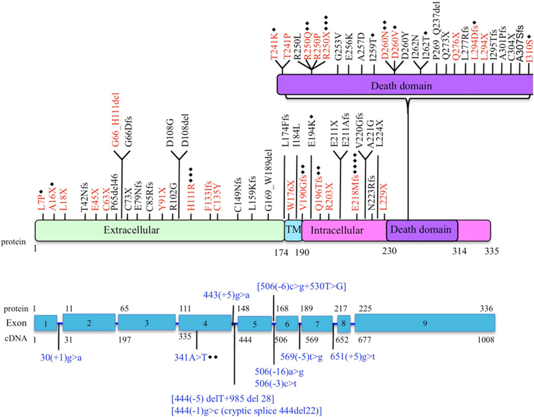
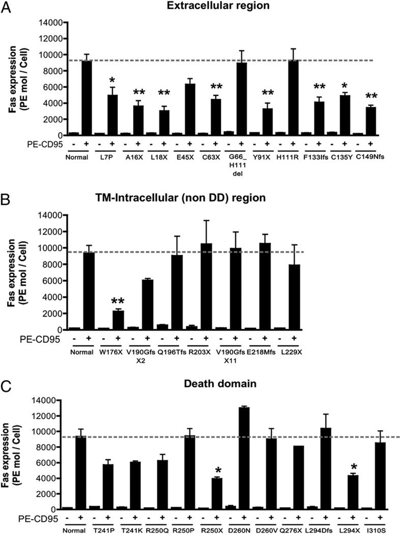
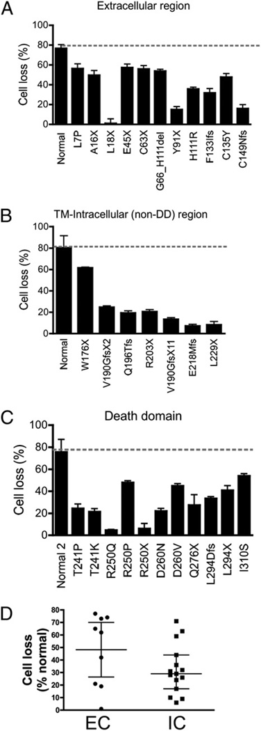
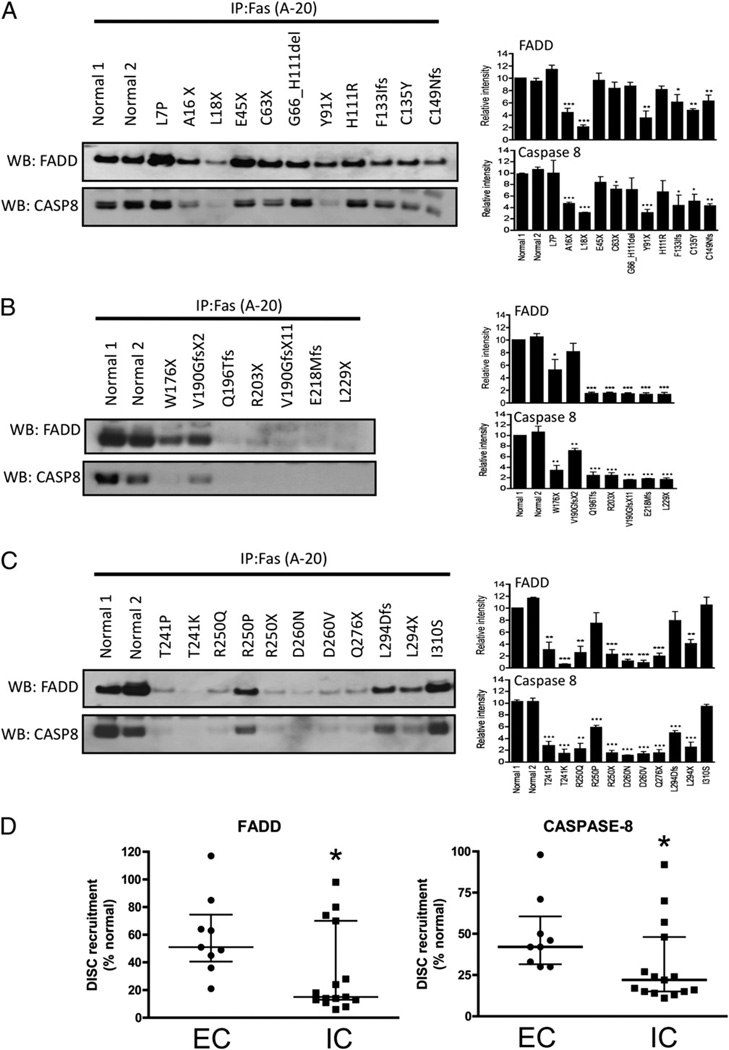
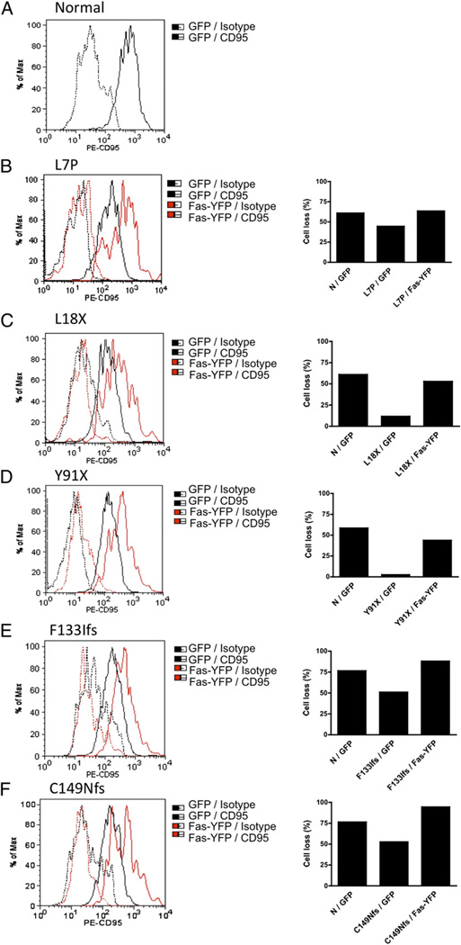
The autoimmune lymphoproliferative syndrome (ALPS) is characterized by early-onset lymphadenopathy, splenomegaly, immune cytopenias, and an increased risk for B cell lymphomas. Most ALPS patients harbor mutations in the FAS gene, which regulates lymphocyte apoptosis. These are commonly missense mutations affecting the intracellular region of the protein and have a dominant-negative effect on the signaling pathway. However, analysis of a large cohort of ALPS patients revealed that ∼30% have mutations affecting the extracellular region of FAS, and among these, 70% are nonsense, splice site, or insertions/deletions with frameshift for which no dominant-negative effect would be expected. We evaluated the latter patients to understand the mechanism(s) by which these mutations disrupted the FAS pathway and resulted in clinical disease. We demonstrated that most extracellular-region FAS mutations induce low FAS expression due to nonsense-mediated RNA decay or protein instability, resulting in defective death-inducing signaling complex formation and impaired apoptosis, although to a lesser extent as compared with intracellular mutations. The apoptosis defect could be corrected by FAS overexpression in vitro. Our findings define haploinsufficiency as a common disease mechanism in ALPS patients with extracellular FAS mutations.


Conflict of interest statement

Disclosures

The authors have no financial conflicts of interest.

Figures

FIGURE 1
FIGURE 1
Schematic representation of FAS mutations in ALPS patients. Red text indicates mutations evaluated in this study. Blue text indicates complex mutations. Black diamonds represent the number of additional families with same mutation. TM, transmembrane.
FIGURE 2
FIGURE 2
Cell-surface expression of FAS on EBV-transformed B cells. A–C, Cells (0.5 × 10⁶) were stained for FAS expression using 10 µg/ml PE-conjugated anti-CD95 or PE-conjugated isotype-matched IgG for 30 min at 4°C. After washing two times with PBS, 10,000 live cells were analyzed by flow cytometry. FAS expression was quantified using QuantiBRITE PE (BD Biosciences). Data were represented as means ± SE of three to four separate experiments. *p < 0.05, **p < 0.01 by Student t test for comparison with normal cell lines.
FIGURE 3
FIGURE 3
Sensitivity of EBV-transformed B cells to CD95-induced cell death. A–C, EBV-transformed B cells from ALPS patients and three different normal controls were stimulated with APO-1–3 (1 µg/ml) and protein A (1 µg/ml) for 24 h. Cell death was determined by measuring the loss of the mitochondrial transmembrane potential using DiOC6 by flow cytometry. D, Comparison of the degree of apoptotic defect between cells with extracellular (EC) mutations (including W176X) affecting FAS expression (n = 9) and intracellular (IC) mutations with normal FAS expression (n = 16), with numbers normalized to the value in normal cells. The data in A–C were represented as means ± SE of three separate experiments. The data in D were presented as medians and interquartile ranges, and medians were compared using Mann–Whitney U test. All results from ALPS patients showed significant reduction in FAS-mediated cell death compared with normal cell lines.
FIGURE 4
FIGURE 4
DISC formation in EBV-transformed B cells of ALPS patients. A–C, Cells were stimulated with APO-1–3 (0.5 µg/ml) or isotype control IgG in the presence of protein A (1 µg/ml) for 20 min. After lysis, cell lysates were incubated with anti-FAS Ab and protein A/G agarose for 2 h. The immunocomplexes were then subjected to Western blotting using anti-FADD and anti–caspase-8 (CASP8). Caspase-8 band represents cleaved caspase-8 (~43 kDa). Because FAS size overlaps with IgG H chain, we could not show immunoprecipitated FAS. D, Comparison of the degree of recruitment of FADD or caspase-8 to the FAS receptor upon stimulation between cells with extracellular mutations affecting FAS expression (EC; n = 9) and intracellular mutations with normal FAS expression (IC; n = 16). All numbers were normalized to the value in normal cells. Densitometry data (A–C) are presented as means ± SE of (n = 3) separate experiments and compared by Student t test. The data in D were presented as medians and interquartile ranges, and medians were compared using Mann–Whitney U test. *p < 0.05, **p < 0.01, ***p , 0.001.
FIGURE 5
FIGURE 5
Overexpression of wild-type FAS into FAS-haploinsufficient samples. A–F, Normal and patient EBV-transformed B cells (3 × 10⁶ cells/sample) were transfected with 5 µg GFP (pEGFP-C1) or 5 µg YFP-FAS (pEYFP-N1-wt FAS) using Amaxa Human B-cell Nucleofector. After 48 h of transfection, over-expressed FAS on the cell surface was determined by flow cytometry using PE-CD95, and cells were stimulated with APO-1–3 (1 µg/ml) and protein A (1 µg/ ml) for 24 h. Cell death was determined by flow cytometry. Analysis was performed by gating on live GFP- or YFP-FAS–transfected cells. Data were presented as means of duplicate experiments. N, normal control.

Similar articles

Cited by

References

    1. Sneller MC, Wang J, Dale JK, Strober W, Middelton LA, Choi Y, Fleisher TA, Lim MS, Jaffe ES, Puck JM, et al. Clincal, immunologic, and genetic features of an autoimmune lymphoproliferative syndrome associated with abnormal lymphocyte apoptosis. Blood. 1997;89:1341–1348. - PubMed
    1. Le Deist F, Emile JF, Rieux-Laucat F, Benkerrou M, Roberts I, Brousse N, Fischer A. Clinical, immunological, and pathological consequences of Fas-deficient conditions. Lancet. 1996;348:719–723. - PubMed
    1. Straus SE, Jaffe ES, Puck JM, Dale JK, Elkon KB, Rösen-Wolff A, Peters AM, Sneller MC, Hallahan CW, Wang J, et al. The development of lymphomas in families with autoimmune lymphoproliferative syndrome with germline Fas mutations and defective lymphocyte apoptosis. Blood. 2001;98:194–200. - PubMed
    1. Sneller MC, Straus SE, Jaffe ES, Jaffe JS, Fleisher TA, Stetler-Stevenson M, Strober W. A novel lymphoproliferative/autoimmune syndrome resembling murine lpr/gld disease. J. Clin. Invest. 1992;90:334–341. - PMC - PubMed
    1. Bleesing JJ, Brown MR, Straus SE, Dale JK, Siegel RM, Johnson M, Lenardo MJ, Puck JM, Fleisher TA. Immunophenotypic profiles in families with autoimmune lymphoproliferative syndrome. Blood. 2001;98:2466–2473. - PubMed

Publication types