. 2011 Apr 13;31(15):5579-88.
doi: 10.1523/JNEUROSCI.6641-10.2011.

Extra forces evoked during electrical stimulation of the muscle or its nerve are generated and modulated by a length-dependent intrinsic property of muscle in humans and cats

Affiliations

Extra forces evoked during electrical stimulation of the muscle or its nerve are generated and modulated by a length-dependent intrinsic property of muscle in humans and cats

Alain Frigon et al. J Neurosci. .

Abstract

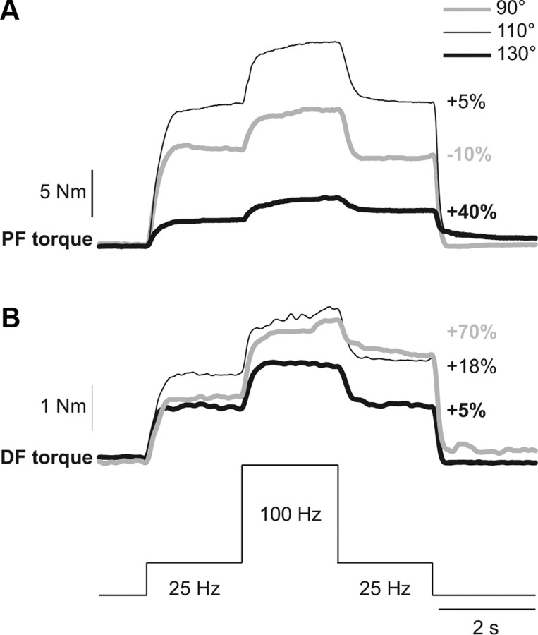
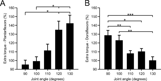
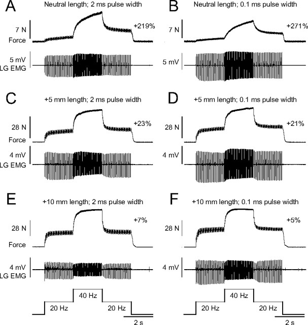
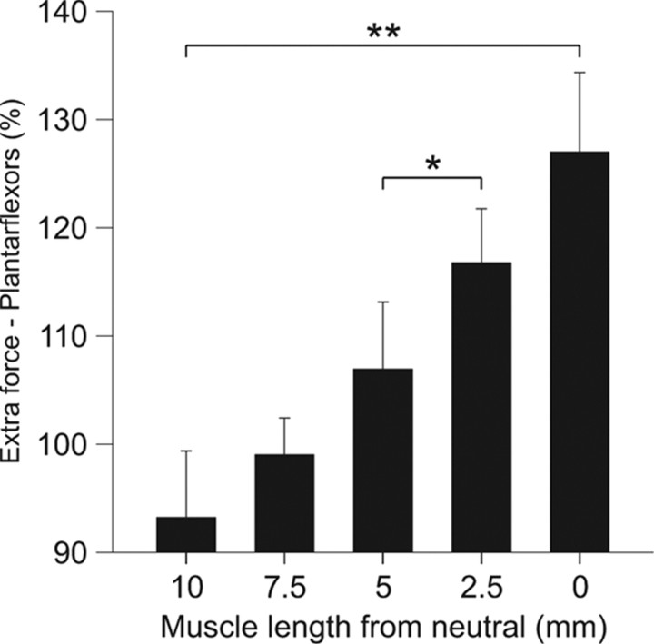
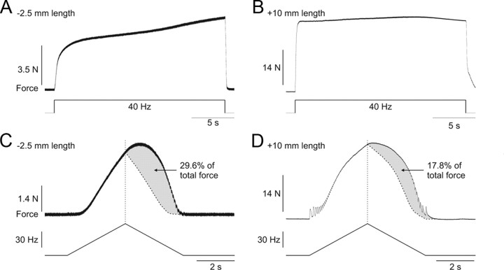
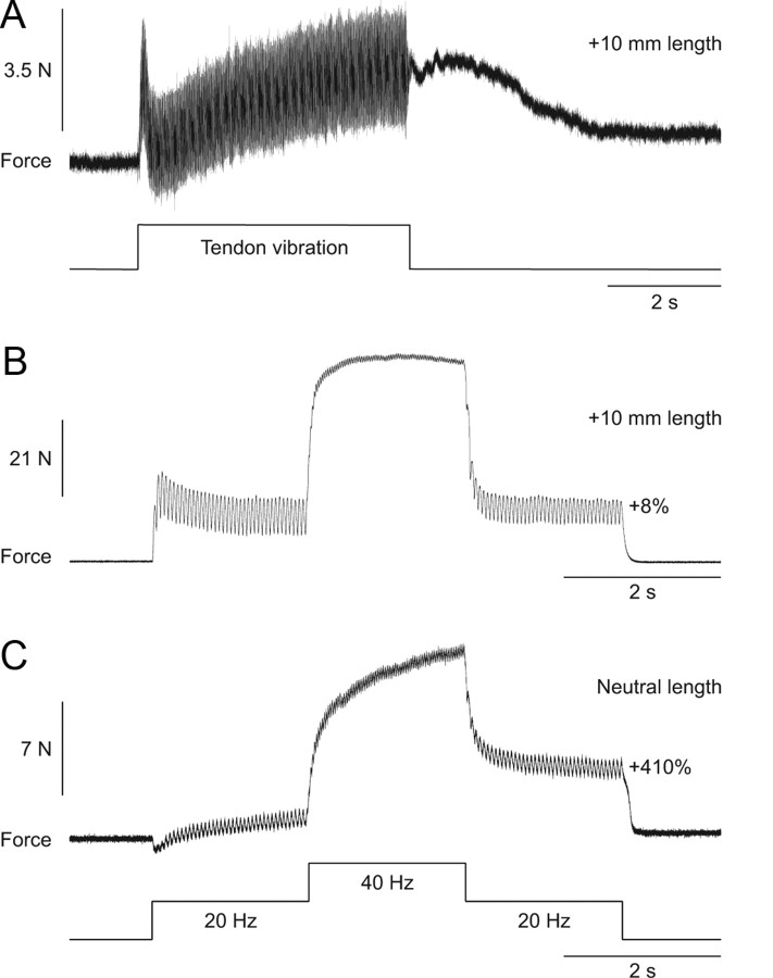
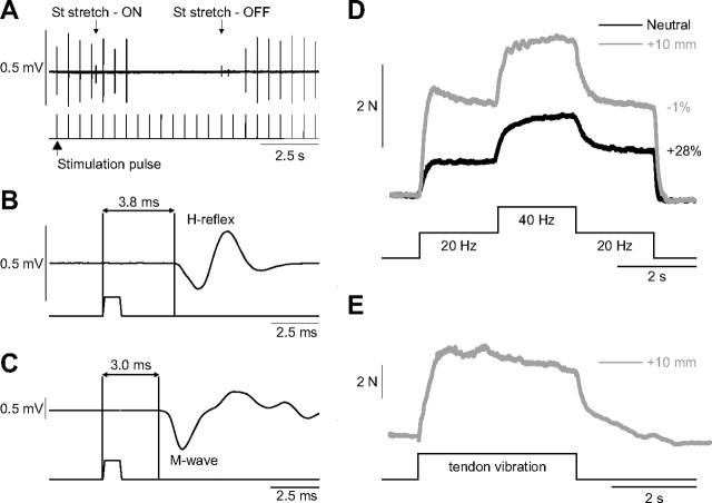
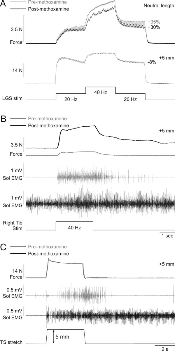
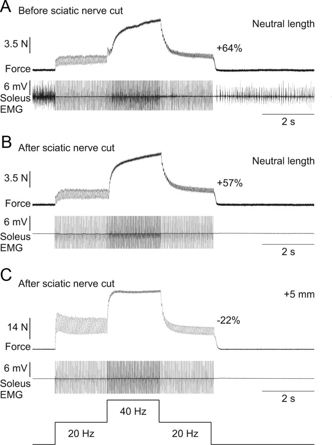
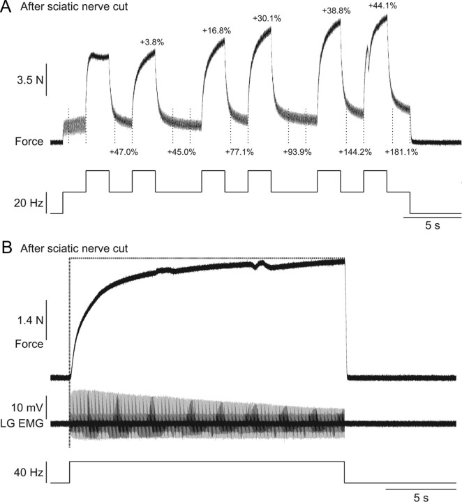
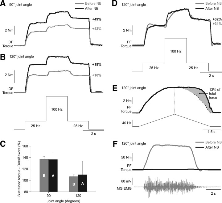
Extra forces or torques are defined as forces or torques that are larger than would be expected from the input or stimuli, which can be mediated by properties intrinsic to motoneurons and/or to the muscle. The purpose of this study was to determine whether extra forces/torques evoked during electrical stimulation of the muscle or its nerve with variable frequency stimulation are modulated by muscle length/joint angle. A secondary aim was to determine whether extra forces/torques are generated by an intrinsic neuronal or muscle property. Experiments were conducted in 14 able-bodied human subjects and in eight adult decerebrate cats. Torque and force were measured in human and cat experiments, respectively. Extra forces/torques were evoked by stimulating muscles with surface electrodes (human experiments) or by stimulating the nerve with cuff electrodes (cat experiments). In humans and cats, extra forces/torques were larger at short muscle lengths, indicating that a similar regulatory mechanism is involved. In decerebrate cats, extra forces and length-dependent modulation were unaffected by intrathecal methoxamine injections, despite evidence of increased spinal excitability, and by transecting the sciatic nerve proximal to the nerve stimulations. Anesthetic nerve block experiments in two human subjects also failed to abolish extra torques and the length-dependent modulation. Therefore, these data indicate that extra forces/torques evoked during electrical stimulation of the muscle or nerve are muscle length-dependent and primarily mediated by an intrinsic muscle property.


Figures

Figure 1.
Figure 1.
Modulation of extra torques at different joint angles in a human subject. Torques evoked by plantarflexor (PF, A) and dorsiflexor (DF, B) top hat stimulations (25, 100, and 25 Hz) at ankle joint angles of 90°, 110°, and 130°. The extra torque (i.e., the torque produced during the second 25 Hz stimulation expressed as a percentage of the first 25 Hz stimulation) is shown on the right for each joint angle.
Figure 2.
Figure 2.
Modulation of extra torques at different joint angles for the group of human subjects. Magnitude of the extra torque (torque evoked during second 25 Hz stimulation ÷ torque evoked during first 25 Hz stimulation × 100) for the group, evoked by plantarflexor (n = 9, A) and dorsiflexor (n = 13, B) top hat stimulations. Asterisks indicate significant differences between joint angles (pairwise comparisons with Bonferroni correction). Each bar is the mean ± SE. *p < 0.05, **p < 0.01, ***p < 0.001.
Figure 3.
Figure 3.
Modulation of extra forces at different joint angles in an adult decerebrate cat. Forces produced by triceps surae muscles evoked by stimulating the LGS nerve during top hat stimulations with pulse widths of 2 ms (A, C, E) or 0.1 ms (B, D, F) at a neutral muscle length (A, B), with the muscle stretched 5 mm (C, D), and 10 mm (E, F). The extra force (i.e., the force produced during the second 20 Hz stimulation expressed as a percentage of the first 20 Hz stimulation) is shown on the right for each muscle length. The EMG of the LG muscle evoked during stimulations is shown below each force trace.
Figure 4.
Figure 4.
Modulation of extra forces at different muscle lengths for the group of adult decerebrate cats. Magnitude of the triceps surae extra forces (force evoked during the second 20 Hz stimulation ÷ the force evoked during first 20 Hz stimulation × 100) for the group (n = 4), evoked by stimulating the LGS or MG nerves with top hat stimulations (20, 40, and 20 Hz). Asterisks indicate significant differences between joint angles (pairwise comparisons with Bonferroni correction). Each bar is the mean ± SE. *p < 0.05, **p < 0.01.
Figure 5.
Figure 5.
Extra forces evoked by other methods and their length-dependent modulation in an adult decerebrate cat. Force produced by stimulating the LGS nerve at 40 Hz for 20 s at muscle lengths of −2.5 mm (A) and +10 mm (B) from neutral. Force produced by stimulating the LGS nerve with a triangular frequency ramp from 4 to 60 Hz and back down to 4 Hz in a 6 s period at muscle lengths of −2.5 mm (C) and +10 mm (D) from neutral. The dotted vertical line represent the point of stimulation at 60 Hz and the curved dotted line represents a smoothed hypothetical symmetric force profile on the descending slope of the frequency ramp. The gray area illustrates the extra force, which is expressed as a percentage of the total force.
Figure 6.
Figure 6.
Extra forces evoked by Achilles tendon vibration and top hat stimulations at two different muscle lengths in an adult decerebrate cat. A, Force output generated by ankle plantarflexors during and following vibration of the Achilles tendon at a muscle length of +10 mm from neutral. B, C, Forces produced during 20, 40, and 20 Hz top hat stimulations at muscle lengths of +10 mm (B) and 0 mm (C) from neutral. The extra force (i.e., the force produced during the second 20 Hz stimulation expressed as a percentage of the first 20 Hz stimulation) during top hat stimulations is shown on the right for each muscle length.
Figure 7.
Figure 7.
Determination of the H-reflex in the VL and extra forces of the quadriceps muscles. A, Stimulation of the Quad nerve in the inguinal region at low stimulation intensities (<1.5 T) evoked compound action potentials in the VL muscle that were abolished by stretching the semitendinosus (St) muscle, thus confirming that the response was an H-reflex. B, Latency of the H-reflex measured from stimulation onset to the first deflection of the EMG trace at a stimulation intensity of 1.1 T. C, Latency of the M-wave, measured from stimulation onset to the first deflection of the EMG trace, at a stimulation intensity of 3.0 T. D, Forces produced with top hat stimulations of the Quad nerve at 1.1 T at muscle lengths +10 and 0 mm from neutral. E, Force output generated by the Quad muscle group during and following vibration of the Quad tendon at a muscle length of +10 mm from neutral.
Figure 8.
Figure 8.
Methoxamine injection increases spinal excitability but has no effect on extra forces in an adult decerebrate cat. A, Forces produced by left triceps surae muscles evoked by stimulating the left LGS nerve during top hat stimulations (Stim, 1 ms pulse widths) before and after injecting methoxamine at neutral and long muscle lengths. B, Force generated by the left triceps surae muscles with stimulation of the right tibial (Tib) nerve (40 Hz for 2 s, 0.2 ms pulse width) before and after injecting methoxamine. C, Ramp-and-hold stretch of the left triceps surae (TS) muscles (ramp: 5 mm in 0.25 s) before and after injecting methoxamine. The EMG of the soleus (Sol) muscle with stimulation of the right tibial nerve (B) and with stretch of triceps surae muscles (C) is shown below the force traces for trials before and after methoxamine injection.
Figure 9.
Figure 9.
Extra forces at short muscle lengths are unchanged following transection of the sciatic nerve in an adult decerebrate cat. A, Force produced by triceps surae muscles evoked by top hat stimulation of the LGS nerve (2 ms pulse widths) before transecting the sciatic nerve with concurrent EMG of the soleus muscle at the neutral muscle length. Note that the soleus is tonically active before and after the top hat stimulation, whereas it is primarily inactive during the stimulation. B, Force produced during top hat stimulation after transecting the sciatic nerve with the same stimulation parameters as in A at the neutral muscle length. C, Force produced during top hat stimulation after transecting the sciatic nerve with the same stimulation parameters as in A at a muscle length of + 5 mm from neutral. Note that soleus EMG is absent in B and C. The extra force (i.e., the force produced during the second 20 Hz stimulation expressed as a percentage of the first 20 Hz stimulation) is shown on the right for each force trace.
Figure 10.
Figure 10.
Progressive increase in extra and peak forces with repeated top hat stimulations and staircase phenomenon after transecting the sciatic nerve in two adult decerebrate cats. A, The LGS nerve was stimulated (2 ms pulse widths) at variable frequencies during a 30 s period after transecting the sciatic nerve. The extra forces (i.e., the forces produced during the last 1.5 s of the 20 Hz stimulations; the period is indicated by vertical dotted lines) and the peak forces produced with each 2 s 40 Hz stimulation expressed as a percentage of first 20 Hz stimulation (i.e., for extra forces) and the first 40 Hz stimulation (i.e., for peak forces) are shown above the trace. B, The LGS nerve was stimulated at 40 Hz for 20 s after transecting the nerve. The EMG of the LG muscle evoked by stimulation is shown below the force trace.
Figure 11.
Figure 11.
Extra torques before and after nerve block in two human subjects. A, B, Torques evoked by dorsiflexor (DF) top hat stimulations (25, 100, and 25 Hz) at ankle joint angles of 90° and 120°, before and after a lidocaine nerve block (NB) of the common peroneal nerve in subject 1. C, Average ± SD of three trials before (B) and after (A) nerve block at ankle joint angles of 90° and 120° for subject 1. D, Torques evoked by plantarflexor (PF) top hat stimulations (25, 100, and 25 Hz) at an ankle joint angle of 120°, before and after a lidocaine nerve block of the tibial nerve in subject 2. E, Torque produced by stimulating the plantarflexor muscles with a triangular frequency ramp from 4 to 80 Hz and back down to 4 Hz in a 6 s period at a joint angle of 120° for subject 2 after nerve block. The dotted vertical line represent the point of stimulation at 80 Hz and the curved dotted line represents a smoothed hypothetical symmetric torque profile on the descending slope of the frequency ramp. The gray area illustrates the extra torque, which is expressed as a percentage of the total torque. F, The torque and EMG of the MG produced during a maximal voluntary effort of the plantarflexors before and after nerve block in subject 2. The percentage increase in the extra torque (i.e., the torque produced during second the 25 Hz stimulation expressed as a percentage of the first 25 Hz stimulation) is shown on the right for each joint angle in A, B, and D.

Similar articles

Cited by

References

    1. Balnave CD, Allen DG. The effect of muscle length on intracellular calcium and force in single fibres from mouse skeletal muscle. J Physiol. 1996;492:705–713. - PMC - PubMed
    1. Banks RW, Barker D, Stacey MJ. Form and distribution of sensory terminals in cat hindlimb muscle spindles. Philos Trans R Soc Lond B Biol Sci. 1982;299:329–364. - PubMed
    1. Bennett DJ, Hultborn H, Fedirchuk B, Gorassini M. Synaptic activation of plateaus in hindlimb motoneurons of decerebrate cats. J Neurophysiol. 1998a;80:2023–2037. - PubMed
    1. Bennett DJ, Hultborn H, Fedirchuk B, Gorassini M. Short-term plasticity in hindlimb motoneurons of decerebrate cats. J Neurophysiol. 1998b;80:2038–2045. - PubMed
    1. Binder-Macleod SA, Barrish WJ. Force response of rat soleus muscle to variable-frequency train stimulation. J Neurophysiol. 1992;68:1068–1078. - PubMed

Publication types

MeSH terms

LinkOut - more resources