. 2011 Apr 13;31(15):5659-72.
doi: 10.1523/JNEUROSCI.6638-10.2011.

Transport of the major myelin proteolipid protein is directed by VAMP3 and VAMP7

Affiliations

Transport of the major myelin proteolipid protein is directed by VAMP3 and VAMP7

Anke Feldmann et al. J Neurosci. .

Abstract

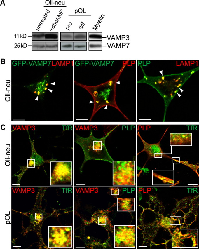
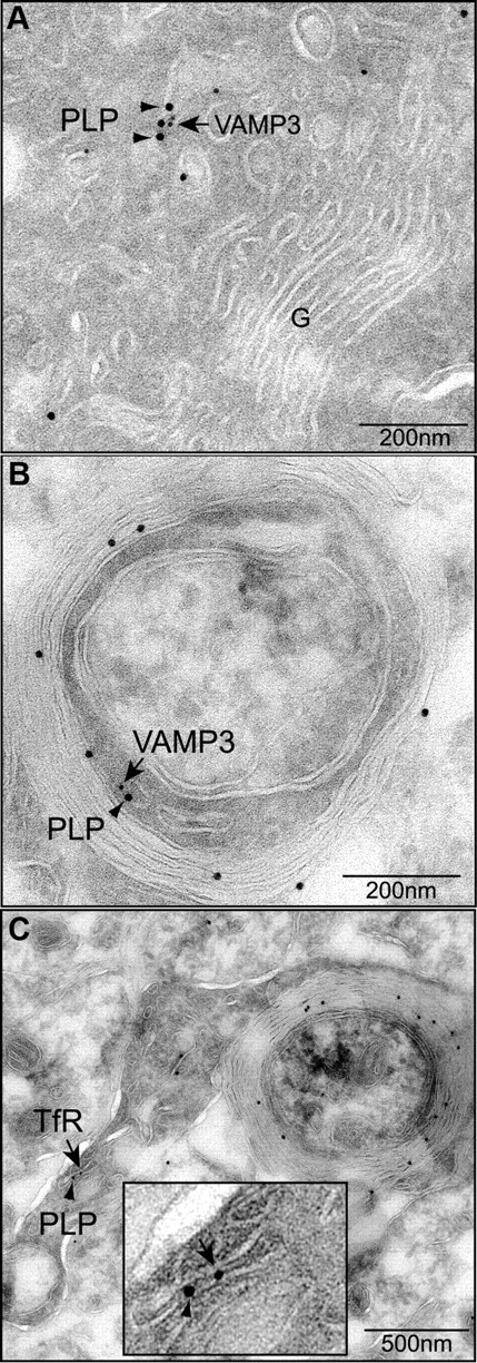
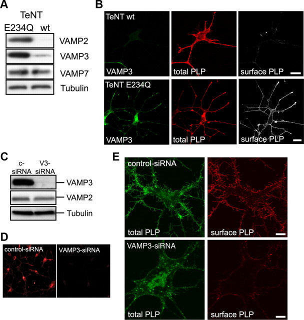
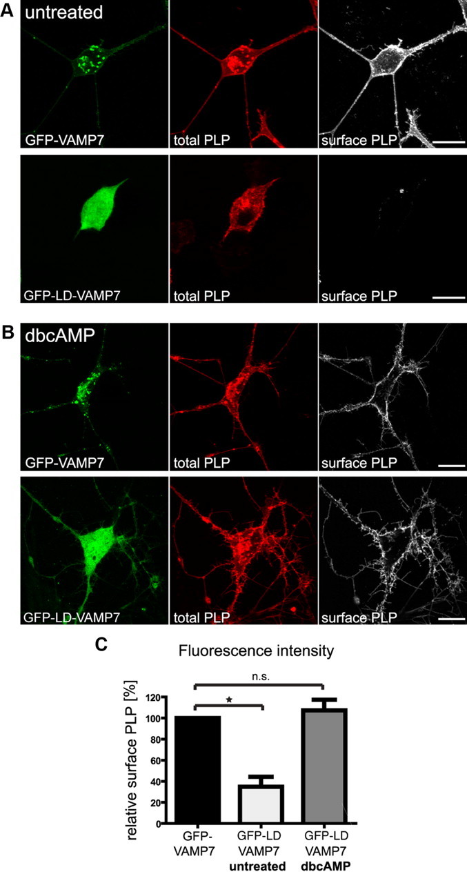
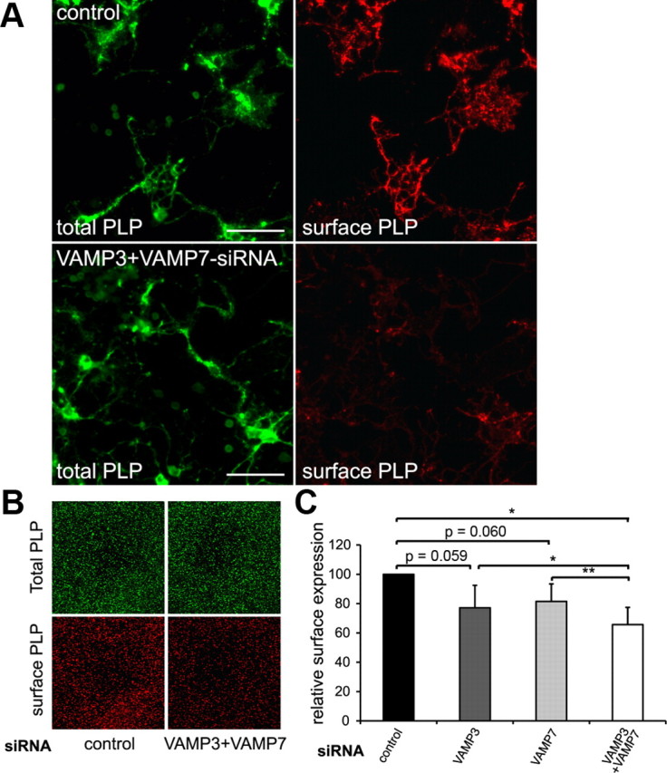
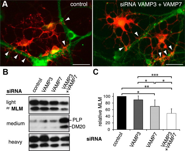
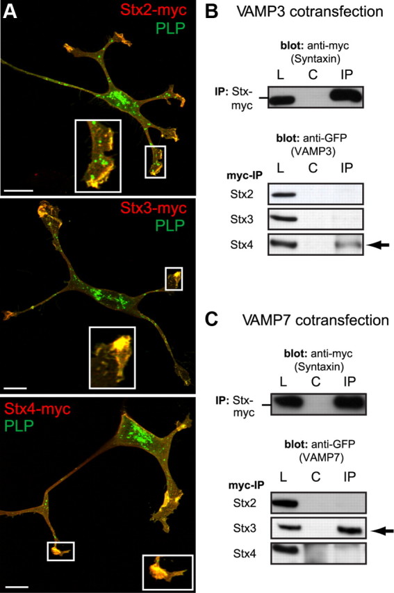
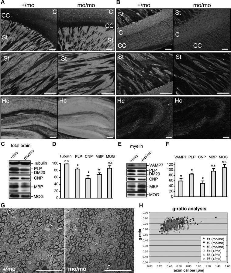
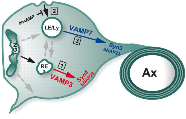
CNS myelination by oligodendrocytes requires directed transport of myelin membrane components and a timely and spatially controlled membrane expansion. In this study, we show the functional involvement of the R-soluble N-ethylmaleimide-sensitive factor attachment protein receptor (R-SNARE) proteins VAMP3/cellubrevin and VAMP7/TI-VAMP in myelin membrane trafficking. VAMP3 and VAMP7 colocalize with the major myelin proteolipid protein (PLP) in recycling endosomes and late endosomes/lysosomes, respectively. Interference with VAMP3 or VAMP7 function using small interfering RNA-mediated silencing and exogenous expression of dominant-negative proteins diminished transport of PLP to the oligodendroglial cell surface. In addition, the association of PLP with myelin-like membranes produced by oligodendrocytes cocultured with cortical neurons was reduced. We furthermore identified Syntaxin-4 and Syntaxin-3 as prime acceptor Q-SNAREs of VAMP3 and VAMP7, respectively. Analysis of VAMP3-deficient mice revealed no myelination defects. Interestingly, AP-3δ-deficient mocha mice, which suffer from impaired secretion of lysosome-related organelles and missorting of VAMP7, exhibit a mild dysmyelination characterized by reduced levels of select myelin proteins, including PLP. We conclude that PLP reaches the cell surface via at least two trafficking pathways with distinct regulations: (1) VAMP3 mediates fusion of recycling endosome-derived vesicles with the oligodendroglial plasma membrane in the course of the secretory pathway; (2) VAMP7 controls exocytosis of PLP from late endosomal/lysosomal organelles as part of a transcytosis pathway. Our in vivo data suggest that exocytosis of lysosome-related organelles controlled by VAMP7 contributes to myelin biogenesis by delivering cargo to the myelin membrane.


Figures

Figure 1.
Figure 1.
Colocalization of VAMP3 and VAMP7 with PLP in endosomal membranes of oligodendroglial cells. A, Western blot analysis of endogenous VAMP3 and VAMP7 protein expression in oligodendroglial cells and myelin isolated from P60 mouse brain. Primary oligodendrocytes (pOL) were analyzed in progenitor (pro) and differentiated (diff) state, cultured for 2 and 8 d, respectively. Oli-neu cells were treated or not for 3 d with dbcAMP. B, Coexpression of GFP-VAMP7 and PLP in Oli-neu cells followed by immunostaining of PLP and LAMP1 reveals colocalization of PLP with GFP-VAMP7 in late endosomes of Oli-neu cells (arrowheads). Note that VAMP7 antibodies failed to detect VAMP7 by immunocytochemical means. C, Coimmunostaining of VAMP3, PLP, and TfR reveals colocalization of PLP with VAMP3 in recycling endosomes of Oli-neu cells or primary oligodendrocytes. Confocal planes are shown; insets show enlarged areas. Scale bars, 5 μm.
Figure 2.
Figure 2.
Ultrastructural analysis of VAMP3 and PLP colocalization. A, B, Immunolabeling of PLP (15 nm gold, arrowheads) and VAMP3 (10 nm gold; arrows) on ultrathin sections of primary oligodendrocytes (A) and myelinating oligodendrocytes (B) of P6 spinal cord. VAMP3 and PLP colocalize on tubular membranes within the oligodendroglial cytoplasm. C, Double labeling of PLP (15 nm gold; arrowheads) and TfR (10 nm gold; arrows) in myelinating oligodendrocytes of P6 spinal cord demonstrates PLP localization to membranes of recycling endosomal identity. The inset depicts enlarged area of colocalization.
Figure 3.
Figure 3.
Functional inactivation of VAMP3-mediated fusion. A, B, Inactivation of VAMP3 by TeNT-mediated proteolytic cleavage. Oli-neu cells were cotransfected with PLP and TeNT light chain (TeNT-wt) or a mutant inactive form (TeNT-E234Q) as control and treated with 1 mm dbcAMP for 3 d. A, Western blot analysis of total cell lysates demonstrating specific cleavage of VAMP2 and VAMP3 but not VAMP7 by TeNT-wt. Tubulin is shown as loading control. B, Confocal imaging of cells stained with antibodies recognizing VAMP3 (green), total PLP (red; aa3 antibody), and surface PLP (white; O10 antibody live stain). TeNT-wt-expressing cells lack VAMP3 and have reduced levels of PLP at their cell surface. Scale bars, 10 μm. C–E, siRNA-mediated silencing of VAMP3 expression in Oli-neu cells stably expressing PLP-EGFP. The cells were transfected with either a nonspecific siRNA (c-siRNA, control) or with VAMP3-siRNA (V3-siRNA) and treated with 1 mm dbcAMP for 3 d. C, Verification of knockdown efficiency and specificity by Western blot analysis of total cell lysates. VAMP2 expression is not affected by the VAMP3-siRNA. Tubulin is shown as loading control. D, Verification of knockdown efficiency by immunocytochemical detection of VAMP3. E, Confocal imaging of control siRNA-treated cells (top panels) and VAMP3 siRNA-treated cells (bottom panels) after treatment for 3 d with dbcAMP showing PLP-EGFP in green (total PLP) and surface PLP in red, as determined by O10 staining.
Figure 4.
Figure 4.
Functional inactivation of VAMP7-mediated fusion by overexpression of the dominant-negative N-terminal Longin domain of VAMP7. Oli-neu cells were transfected to coexpress PLP and EGFP fused to full-length VAMP7 (GFP-VAMP7) or the Longin-domain of VAMP7 (GFP-LD-VAMP7). A, B, Immunostaining of cells using antibodies recognizing total PLP (aa3 antibody; red) or surface PLP (O10 antibody; white). PLP surface localization is strongly reduced in untreated cells (A) but unaffected in dbcAMP-treated cells (B). Scale bars, 10 μm. C, Relative levels of surface PLP calculated by quantification of fluorescence intensity from confocal image stacks of dbcAMP-treated or untreated cells (O10 staining intensity of surface PLP in relation to aa3 staining intensity of total PLP). Error bars depict SEM. Paired t test, *p < 0.05; n.s., not significant; n = 3.
Figure 5.
Figure 5.
VAMP3 and VAMP7 silencing in primary oligodendrocytes. OPCs were siRNA transfected and differentiated for 3–4 d to allow endogenous PLP expression. A, Surface-localized and total PLP were determined by immunostaining using O10 and aa3 antibodies, respectively. Scale bars, 50 μm. B, Odyssey infrared scan depicting total PLP and surface PLP of control siRNA-transfected and VAMP3/VAMP7-cosilenced oligodendrocytes. A detail showing an identical field of the scan is shown. C, Surface PLP normalized to total PLP calculated from Odyssey scans of siRNA-treated cells (n = 5 for single and n = 4 for double silencing experiments). Error bars depict SEM. Paired t test, *p < 0.05, **p < 0.01.
Figure 6.
Figure 6.
VAMP3 and VAMP7 silencing in neuron–oligodendrocyte cocultures. siRNA-transfected primary oligodendrocytes were seeded on top of primary cortical neurons and cultured for an additional 5 d to allow initial axon–glia interaction and ensheathment of axons with MLMs. A, Immunostaining of cocultures using antibodies recognizing βIII-tubulin as neuronal (green) and PLP as oligodendroglial marker (red). Morphology of oligodendrocytes and extent of axon–glia interaction appeared similar between control cultures and the different siRNA treatment conditions. The arrowheads mark sites of glial axon ensheathment. Scale bars, 25 μm. B, C, Cocultures were subjected to gradient centrifugation to isolate membrane fractions of high-, medium-, and low-density (light fraction). Light fractions contain MLMs sharing the characteristics of myelin in vivo. B, Western blot showing the distribution of PLP between light, medium, and heavy fractions under the different conditions of siRNA treatment. C, Densitometric quantification of the relative association of PLP with MLMs (n = 5). On RNA silencing of VAMP7 and cosilencing of VAMP3 and VAMP7, association of PLP with the MLM fraction is significantly reduced. Error bars depict SEM. Paired t test, *p < 0.05, **p < 0.01, ***p < 0.001.
Figure 7.
Figure 7.
The Qa-SNAREs Stx3 and Stx4 function as putative target receptors of VAMP3 and VAMP7 in oligodendroglial cells. A, Confocal images of Oli-neu cells cotransfected with PLP and myc-tagged versions of Stx2, Stx3, or Stx4. Immunostainings were performed using aa3 antibodies to detect total PLP (green) and myc antibodies to detect syntaxins (red). Colocalization appears prominently in clusters at terminal ends of the processes (enlarged view in insets). B, C, IP of myc-tagged Stx2, Stx3, or Stx4 from lysates of cells cotransfected with GFP-VAMP3 (B) or GFP-VAMP7 (C) using polyclonal myc antibodies. IPs were analyzed by Western blotting using either monoclonal myc antibodies to confirm syntaxin precipitation (top panel) or anti-GFP antibodies detecting coprecipitating VAMP3 or VAMP7 (bottom panels). GFP-VAMP3 is selectively detected in IPs of Stx4-myc, and GFP-VAMP7 is selectively detected in IPs of Stx3myc (arrows). L, Total lysates; C, negative controls showing precipitations without primary antibody included; IP, immunoprecipitation.
Figure 8.
Figure 8.
Analysis of myelination in AP-3δ-deficient mocha mice. A, B, Gallyas staining (A) and immunohistochemical staining (B) of PLP of white matter tracts in brains of P30 mocha (mo/mo) and heterozygous (+/mo) littermate control mice. C, Cortex; CC, corpus callosum; St, striatum; Cb, cerebellum; Hc, hippocampus. Scale bars, 100 μm. C, D, Western blot analysis of myelin proteins in brain homogenates from P30 mocha and heterozygous littermate control mice normalized to total protein. Tubulin is shown as loading control. D, Densitometric quantification of Western blots depicting relative levels of myelin proteins in mocha brains normalized to heterozygous littermates (n = 3; SEM; paired t test, *p < 0.05). E, F, Western blot analysis of isolated myelin from P30 mocha and heterozygous littermate control mice normalized to total myelin protein. F, Densitometric quantification of Western blots showing relative levels of proteins in mocha myelin normalized to heterozygous littermates (n = 3; SEM; paired t test, *p < 0.05). G, Electron micrographs taken from the region of the corpus callosum of mocha mice (mo/mo) and heterozygous littermates (+/mo). Scale bar, 5 μm. H, g ratio analysis of myelin sheath thickness in relation to axon diameter in mocha mice.
Figure 9.
Figure 9.
Model of VAMP3- and VAMP7-dependent surface transport. VAMP3 and VAMP7 control two distinct transport pathways of PLP. VAMP3 acts as R-SNARE mediating fusion of PLP-containing vesicles traveling from RE to the plasma membrane (1). Stx4 and SNAP23 may serve as cognate target Q-SNARES for membrane fusion in this pathway. On endocytosis, PLP travels to LE/Lys (2). VAMP7 mediates fusion in exocytic trafficking of PLP from LE/Lys implicated in myelin formation (3). Stx3 and SNAP23 are putative target SNAREs of the VAMP7-dependent pathway. The mode of PLP trafficking is influenced by cAMP signaling. Elevated cAMP results in depletion of LE/Lys pools because of inhibition of endocytosis. Under these conditions, the VAMP3-dependent pathway is prevailing.

References

    1. Advani RJ, Yang B, Prekeris R, Lee KC, Klumperman J, Scheller RH. VAMP-7 mediates vesicular transport from endosomes to lysosomes. J Cell Biol. 1999;146:765–776. - PMC - PubMed
    1. Ang AL, Taguchi T, Francis S, Fölsch H, Murrells LJ, Pypaert M, Warren G, Mellman I. Recycling endosomes can serve as intermediates during transport from the Golgi to the plasma membrane of MDCK cells. J Cell Biol. 2004;167:531–543. - PMC - PubMed
    1. Arantes RM, Andrews NW. A role for synaptotagmin VII-regulated exocytosis of lysosomes in neurite outgrowth from primary sympathetic neurons. J Neurosci. 2006;26:4630–4637. - PMC - PubMed
    1. Baron W, Hoekstra D. On the biogenesis of myelin membranes: sorting, trafficking and cell polarity. FEBS Lett. 2010;584:1760–1770. - PubMed
    1. Bock JB, Matern HT, Peden AA, Scheller RH. A genomic perspective on membrane compartment organization. Nature. 2001;409:839–841. - PubMed

Publication types

MeSH terms

Substances

LinkOut - more resources