. 2011 Apr 13;31(15):5673-81.
doi: 10.1523/JNEUROSCI.0394-11.2011.

Transcription factor Foxd1 is required for the specification of the temporal retina in mammals

Affiliations

Transcription factor Foxd1 is required for the specification of the temporal retina in mammals

Maria Isabel Carreres et al. J Neurosci. .

Abstract

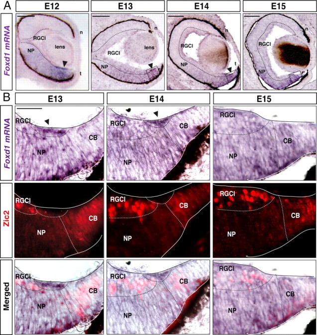
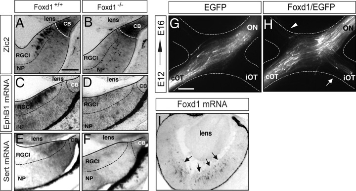
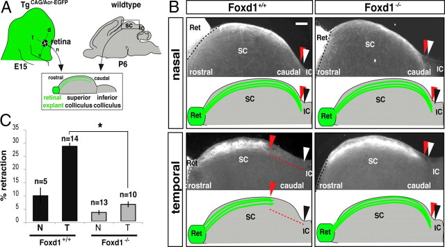
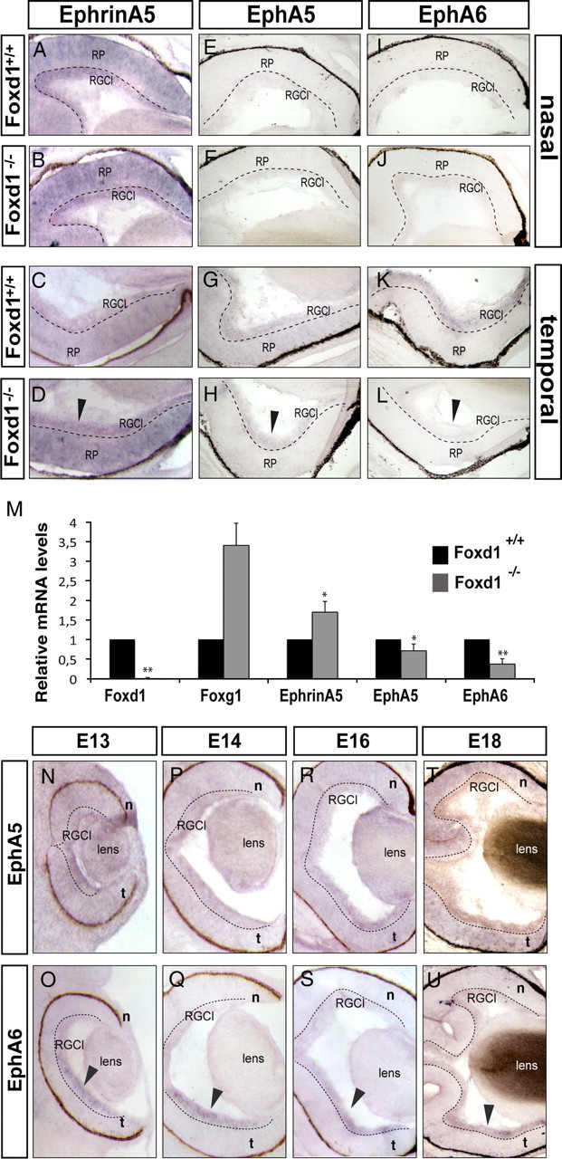
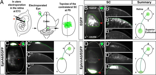
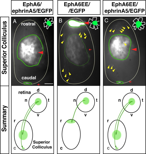
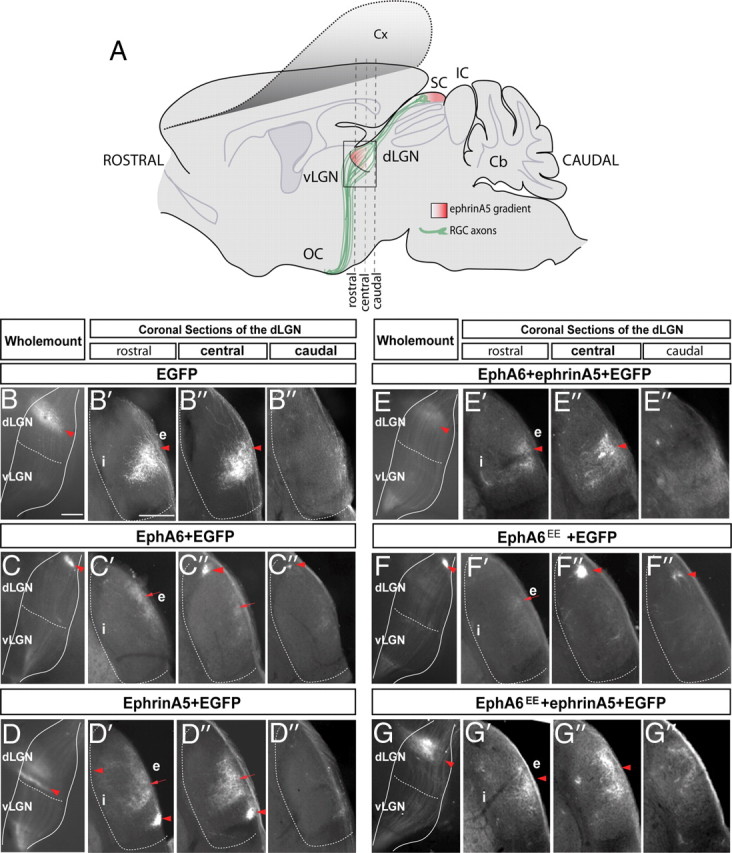
The organization of the visual system is different in birds and mammals. In both, retinal axons project topographically to the visual targets in the brain; but whereas in birds visual fibers from the entire retina decussate at the optic chiasm, in mammals, a number of axons from the temporal retina diverge at the midline to project ipsilaterally. Gain-of-function experiments in chick raised the hypothesis that the transcription factor Foxd1 specifies retinal temporal identity. However, it remains unknown whether Foxd1 is necessary for this function. In mammals, the crucial role of Foxd1 in the patterning of the optic chiasm region has complicated the interpretation of its cell-autonomous function in the retina. Furthermore, target molecules identified for Foxd1 are different in chicks and mice, leading to question the function of Foxd1 in mammals. Here we show that in the mouse, Foxd1 imprints temporal features in the retina such as axonal ipsilaterality and rostral targeting in collicular areas and that EphA6 is a Foxd1 downstream effector that sends temporal axons to the rostral colliculus. In addition, our data support a model in which the desensitization of EphA6 by ephrinA5 in cis is not necessary for the proper functioning of EphA6. Overall, these results indicate that Foxd1 functions as a conserved determinant of temporal identity but reveal that the downstream effectors, and likely their mechanisms of action, are different in mammals and birds.


Figures

Figure 1.
Figure 1.
Spatiotemporal expression of Foxd1 in the developing retina. A, In situ hybridization for Foxd1 in horizontal retinal sections from E12, E13, E14, and E15 wild-type mice shows that in addition to being expressed at low levels in the temporal retina, Foxd1 mRNA is highly accumulated in a peripheral region of the VT retina (arrow). B, The images show combined in situ hybridization for Foxd1 (gray) in retinal sections from the VT retina and immunofluorescence for Zic2 (red) at the indicated stages. Note that an accumulation of Foxd1 mRNA (arrows) between the two Zic2-positive populations, the ciliary body cells, and the iRGCs is appreciable. By E15, Foxd1 mRNA expression in the peripheral region of the VT retina has nearly disappeared. NP, Neural progenitors; n, nasal; t, temporal. RGCl, retinal ganglion cells layer; CB, ciliary body. Scale bars: A, 100 μm; B, 200 μm.
Figure 2.
Figure 2.
Foxd1 nourishes iRGC identity. A, B, Immunohistochemistry against Zic2 in E16 retinal sections from the VT region of wild-type and littermate Foxd1 null embryos shows that although Zic2 expression is normal in the ciliary body cells, it is not detected in differentiated iRGCs in Foxd1-deficient retinas (Foxd1−/−). C–F, In situ hybridization for EphB1 (C, D) or Sert (E, F) in retinal sections from wild-type and Foxd1−/− mice shows that the expression of these two molecules is highly downregulated in VT retina in the absence of Foxd1. NP, Neural progenitors; RGCl, retinal ganglion cells layer; CB, ciliary body. Scale bar, 200 μm. G, H, Images show the chiasm region of E16 embryos electroporated with EGFP or Foxd1/EGFP at E12. ON, Optic nerve; iOT, ipsilateral optic tract; cOT, contralateral optic tract. The white arrow in H points to the ectopic ipsilateral projection. The arrowhead points to misrouted axons in the contralateral optic nerve. Scale bar, 200 μm. I, In situ hybridization to Foxd1 in a retinal section from an E16 embryo electroporated with Foxd1/EGFP confirms ectopic expression of Foxd1 in the central retina (arrows).
Figure 3.
Figure 3.
Temporal axons from Foxd1 null embryos project aberrantly to caudal areas in the SC. A, Schema representing the experimental procedure in which EGFP-labeled retinal explants from wild-type or Foxd1 mutant mice are confronted with wild-type collicular slices. B, Cocultures of nasal or temporal retinal explants from E14 Foxd1+/+;TgCAG/Acr—EGFP or Foxd1−/−;TgCAG/Acr—EGFP embryos confronted to collicular sagittal slices from P6 wild-type mice. Retinal axons from nasal regions of Foxd1−/−;TgCAG/Acr—EGFP mice or from Foxd1+/+;TgCAG/Acr—EGFP reach the end of the colliculus. Temporal axons from Foxd1+/+;TgCAG/Acr—EGFP mice project to mediorostral collicular positions, whereas temporal axons from Foxd1lacZ/lacZ;TgCAG/Acr—EGFP project to caudal areas. White arrowheads mark the end of the SC, and gray arrowheads indicate the location where most of retinal axons stop their growth. Ret, Retinal explant; IC, inferior colliculus. Red dashed lines highlight the long distance between the white and the gray arrowhead in the control situation. Scale bar, 100 μm. C, Quantification of the coculture experiments shows that the aberrant behavior of temporal axons in the absence of Foxd1 is significantly different from wild-type temporal axons. T, temporal; N, Nasal. The values represent means ± SEM (error bars) of the number of cocultures indicated in each case (*p < 0.05, Student's unpaired t test).
Figure 4.
Figure 4.
Expression of EphAs and ephrinAs in wild-type and Foxd1 mutant retinas. A–L, Images show in situ hybridization using specific probes for ephrinA5, EphA5, and EphA6 in horizontal retinal sections from E17 wild-type and Foxd1−/− embryos. The expression of these molecules in the nasal retina of Foxd1−/− mice is unaltered. In contrast, arrows in D, H, and L highlight the altered expression of ephrinA5, EphA5, and EphA6 in the temporal retina of Foxd1−/− mice. M, The graph shows the results from qRT-PCR assays comparing mRNA levels of Foxd1, EphA5, EphA6, Foxg1, and EphrinA5 in E16 retinas from Foxd1+/+ and Foxd1−/− embryos. In the absence of Foxd1, global levels of EphA5 and EphA6 mRNAs decrease, whereas ephrinA5 and Foxg1 mRNA levels increase. mRNA levels for each molecule are shown relative to the wild-type values. At least two retinas for each condition were pooled per experiment, and the average of seven experiments is shown. Error bars indicate ± SEM (*p < 0.05; **p ≤ 0.001, Student's unpaired t test). N–T, In situ hybridization for EphA5 in horizontal retinal sections from embryos at the indicated stages. O–U, In situ hybridization for EphA6 in horizontal retinal sections from embryos at the indicated stages. Arrows point to the strong expression of EphA6 in the temporal RGC layer (RGCl).
Figure 5.
Figure 5.
Ectopic expression of EphA6/EGFP or ephrinA5/EGFP sends retinal axons to the rostral and to the caudal colliculus, respectively. A, Scheme summarizing the experimental procedure. Different combinations of plasmids were injected into the retinas of E13 embryos, and each embryo was then electroporated. Two weeks later, the cortex was removed, and the entire SC was exposed for visualization of EGFP-expressing axons. B–D, The images show a top view of the entire SC of P9 mice electroporated at E13 with the plasmids indicated in each case. In the right corner of each panel, a drawing of the retina to indicate the location of targeted cells (green) is shown. The red arrowheads highlight the termination zone (TZ) of the majority of the targeted axons. Red arrows point the TZ of a minority of the targeted axons. Middle, Coronal sections at the levels indicated by dashed lines. Right, Schemes summarize the projection patterns of targeted RGCs. d, Dorsal; v, ventral; t, temporal; n, nasal; r, rostral; c, caudal. Scale bar, 200 μm.
Figure 6.
Figure 6.
EphrinA5 acts as a receptor in the retina rather than a modifier of the EphA signaling. The images show top views of the SC of P9 mice electroporated at E13 with EphA6/ephrinA5/EGFP (A), EphA6EE/EGFP (B), or EphA6EE/ephrinA5/EGFP (C). Red arrowheads highlight the termination zone (TZ) of the majority of the targeted axons. Red arrows indicate the TZ of a minority of targeted axons. Yellow arrowheads point to axons that seem to be degenerating. In the right corner of each panel, a drawing of the retina to indicate the location of targeted cells (green) is shown. Below each picture, schemes summarize the projection patterns of targeted RGCs in the SC. d, Dorsal; v, ventral; t, temporal; n, nasal; r, rostral; c, caudal. Scale bar, 200 μm.
Figure 7.
Figure 7.
The EphA/ephrinA mechanisms that control the retinothalamic projection are similar than those controlling the retinocollicular projection. A, Schematic representation of a lateral view of the brain in which, after cortex removal, it is possible to observe electroporated retinal axons (green) at the LGN level (squared area). The gradient of ephrinA5 in the dLGN and SC is represented in red. B–G, Lateral view of the entire LGN of P9 mice that were electroporated at E13 with the plasmids indicated in each case. At the right of the images depicting the entire LGNs, coronal sections of the rostral, central, and caudal regions of each dLGN are shown. Red arrowheads highlight the termination zone (TZ) of the majority of the targeted axons. Red arrows indicate the TZ of a minority of the targeted axons. Cx, Cortex; SC superior colliculus; IC, inferior colliculus; vLGN, ventral lateral geniculate nucleus; dLGN, dorsal geniculate nucleus; Cb, cerebellum; OC, optic chiasm; i, internal region of the dLGN; e, external region of the dLGN. Scale bar, 200 μm.

References

    1. Bardelli A, Longati P, Gramaglia D, Basilico C, Tamagnone L, Giordano S, Ballinari D, Michieli P, Comoglio PM. Uncoupling signal transducers from oncogenic MET mutants abrogates cell transformation and inhibits invasive growth. Proc Natl Acad Sci U S A. 1998;95:14379–14383. - PMC - PubMed
    1. Brown A, Yates PA, Burrola P, Ortuno D, Vaidya A, Jessell TM, Pfaff SL, O'Leary DD, Lemke G. Topographic mapping from the retina to the midbrain is controlled by relative but not absolute levels of EphA receptor signaling. Cell. 2000;102:77–88. - PubMed
    1. Brown LY, Kottman AH, Brown S. Immunolocalization of zic2 expression in the developing forebrain. Gene Expr Patterns. 2003;3:361–367. - PubMed
    1. Carvalho RF, Beutler M, Marler KJ, Knoll B, Becker-Barroso E, Heintzmann R, Ng T, Drescher U. Silencing of EphA3 through a cis interaction with ephrinA5. Nat Neurosci. 2006;9:322–330. - PubMed
    1. Chen LI, Webster MK, Meyer AN, Donoghue DJ. Transmembrane domain sequence requirements for activation of the p185c-neu receptor tyrosine kinase. J Cell Biol. 1997;137:619–631. - PMC - PubMed

Publication types

MeSH terms