. 2011 Apr 13;31(15):5699-709.
doi: 10.1523/JNEUROSCI.3477-10.2011.

Neuregulin-1 signals from the periphery regulate AMPA receptor sensitivity and expression in GABAergic interneurons in developing neocortex

Affiliations

Neuregulin-1 signals from the periphery regulate AMPA receptor sensitivity and expression in GABAergic interneurons in developing neocortex

Yuichi Abe et al. J Neurosci. .

Abstract

Neuregulin-1 (NRG1) signaling is thought to contribute to both neuronal development and schizophrenia neuropathology. Here, we describe the developmental effects of excessive peripheral NRG1 signals on synaptic activity and AMPA receptor expression of GABAergic interneurons in postnatal rodent neocortex. A core peptide common to all NRG1 variants (eNRG1) was subcutaneously administered to mouse pups. Injected eNRG1 penetrated the blood-brain barrier and activated ErbB4 NRG1 receptors in the neocortex, in which ErbB4 mRNA is predominantly expressed by parvalbumin-positive GABAergic interneurons. We prepared neocortical slices from juvenile mice that were receiving eNRG1 subchronically and recorded inhibitory synaptic activity from layer V pyramidal neurons. Postnatal eNRG1 treatment significantly enhanced polysynaptic IPSCs, although monosynaptic IPSCs were not affected. Examination of excitatory inputs to parvalbumin-containing GABAergic interneurons revealed that eNRG1 treatment significantly increased AMPA-triggered inward currents and the amplitudes and frequencies of miniature EPSCs (mEPSCs). Similar effects on mEPSCs were observed in mice treated with a soluble, full-length form of NRG1 type I. Consistent with the electrophysiologic data, expression of the AMPA receptor GluA1 (i.e., GluR1, GluRA) was upregulated in the postsynaptic density/cytoskeletal fraction prepared from eNRG1-treated mouse neocortices. Cortical GABAergic neurons cultured with eNRG1 exhibited a significant increase in surface GluA1 immunoreactivity at putative synaptic sites on their dendrites. These results indicate that NRG1 circulating in the periphery influences postnatal development of synaptic AMPA receptor expression in cortical GABAergic interneurons and may play a role in conditions characterized by GABA-associated neuropathologic processes.


Figures

Figure 1.
Figure 1.
Subcutaneous eNRG1 injection activates ErbB4 receptors in the frontal cortex of postnatal mice. A, The time course of ErbB4 phosphorylation was examined after eNRG1 injections in neonatal mice (P2). Cortical tissues were removed at 0.5, 1, 3, 6, 12, and 24 h after injection and subjected to immunoblotting with anti-phospho-ErbB4 or anti-ErbB4 antibodies. B, Protein levels of ErbB3 and ErbB4 in the frontal cortex were examined at P0, P3, P10, P18, and P60. Phosphorylation levels of ErbB3 and ErbB4 were compared at 0, 1, and 3 h after injection. Note that ErbB3 levels in early postnatal days were very limited. C, Postnatal BBB penetration by eNRG1 was also estimated by monitoring ErbB4 phosphorylation at P2, P5, P8, and P11. C, Control; N, eNRG1. D, The distribution of ErbB4 mRNA was examined in the frontal cortices of juvenile mice using in situ hybridization and immunohistochemistry. Most ErbB4 mRNA colocalized with parvalbumin in frontal cortical layer V at P18, as revealed by double staining for parvalbumin (green) and ErbB4 mRNA (red). Note that the frequency of parvalbumin-containing neurons was calculated from a total of 10,608 cells expressing ErbB4 mRNA and that of ErbB4 mRNA-containing neurons was from a total of 5939 parvalbumin-positive cells expressing ErbB4 mRNA in the whole cortex (both 23 sections from four mice).
Figure 2.
Figure 2.
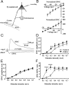
Polysynaptic IPSCs recorded in cortical pyramidal neurons of postnatal mice treated with eNRG1. A, A schematic of the experimental circuit used for patch-clamp recordings shown in C–F. Slices of frontal cortex were prepared from the P11–P12 postnatal mice that had been treated with eNRG1 or saline for 9 d. Recordings were made from layer V pyramidal neurons, and electrical stimuli were delivered to the white matter (WM). B, Estimation of reversal potentials in evoked EPSCs and IPSCs. Normalized I–V curves for EPSCs and IPSCs in layer V pyramidal neurons were plotted. Synaptic currents were evoked by layer VI stimulation. EPSCs were isolated using 10 μm bicuculline and 25 μm APV. IPSCs were isolated using 10 μm CNQX and 25 μm APV in the bath. The mean reversal potentials for EPSCs and IPSCs were estimated to be +4 and −47.5 mV, respectively (n = 4 for each). C, Typical traces for IPSCs and EPSCs are displayed. EPSCs were isolated at a holding potential of −47.5 mV. IPSCs were isolated at a holding potential of +4 mV. Of note, IPSCs with delayed onset latencies appeared to be evoked polysynaptically, because all of these IPSC components were blocked by CNQX. D, E, Polysynaptic IPSCs (D) and EPSCs (E) were recorded from pyramidal neurons in the presence of 25 μm APV. F, The IPSC/EPSC ratio was calculated for each neuron and plotted (control, n = 14 cells; NRG1, n = 17 cells). *p < 0.05, **p < 0.01, ***p < 0.001 compared with control mice at each stimulus intensity by Fisher's LSD.
Figure 3.
Figure 3.
Monosynaptic IPSCs recorded in pyramidal neurons from mice treated with eNRG1. A, A schematic of the experimental circuit. Recordings were made from layer V pyramidal neurons responding to local stimuli delivered 100–200 μm away from the recording electrode. B, IPSCs were recorded at a holding potential of +4 mV in the presence of 10 μm CNQX and 25 μm APV (control, n = 23 cells; eNRG1, n = 26 cells). Of note, we assumed that the CNQX-resistant IPSCs were monosynaptic. C, Typical traces show IPSCs elicited by a paired pulse in control and eNRG1-treated mice. D, The paired-pulse ratios (peak amplitude of EPSC2/peak amplitude of EPSC1) at each stimulus interval (25, 50, 100, and 200 ms) were calculated (control, n = 11 cells; eNRG1, n = 17 cells).
Figure 4.
Figure 4.
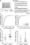
Amplitudes and frequencies of mEPSCs in fast-spiking neurons in postnatal mouse neocortex. A, Putative parvalbumin-positive interneurons were identified based on characteristic firing properties, i.e., nonadapting repetitive firing of fast spikes at >70 Hz with an 800 pA current injection. B, Typical traces of mEPSCs from eNRG1-treated and vehicle-treated (control) mice. C, Cumulative distributions of mEPSC amplitudes and interevent intervals were calculated from spontaneous synaptic events recorded from fast-spiking neurons from layer V. D, Mean amplitudes (left) and frequencies (right) of mEPSCs in the same cells (control, n = 20 cells; eNRG1, n = 20 cells). **p < 0.01, Mann–Whitney U test.
Figure 5.
Figure 5.
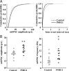
Subchronic effects of fNRG1 on excitatory transmission in fast-spiking neurons. Slices of frontal cortex were prepared from the P11–P12 postnatal mice that had been treated with fNRG1 for 9 d. A, Cumulative distributions of mEPSC amplitudes and interevent intervals were calculated from spontaneous synaptic events recorded from fast-spiking neurons from layer V. B, Mean mEPSC amplitudes (left) and frequencies (right) in the same cells (control, n = 14 cells; eNRG1, n = 12 cells). *p < 0.05, **p < 0.01, Mann–Whitney U test.
Figure 6.
Figure 6.
Delayed influences of postnatal eNRG1 treatment on frequencies of mEPSCs in fast-spiking neurons. Newborn mice were treated with eNRG1 for 9 d as described in Figure 2, but brain slices were prepared at P21–P23. A, Cells were labeled with biocytin and immunostained with anti-parvalbumin antibody (green) and Alexa Flour 594-conjugated streptavidin (red) after they were recorded. Of note, the strong fluorescent signals from the soma produced a flare artifact, which enlarges the soma appearance in these images. In comparison, parvalbumin immunoreactivity in the soma (yellow) is not so strong and looks relatively smaller. Scale bar, 50 μm. B, Cumulative distributions of mEPSC amplitudes and interevent intervals were calculated from spontaneous synaptic events recorded from fast-spiking neurons from layer V. C, Mean mEPSC amplitudes (left) and frequencies (right) in the same cells (control, n = 23 cells; eNRG1, n = 23 cells). *p < 0.05, Mann–Whitney U test.
Figure 7.
Figure 7.
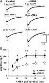
AMPA-evoked inward currents in fast-spiking neurons in eNRG1-treated and vehicle-treated mice. Slices of frontal cortex were prepared from the P11–P12 postnatal mice that had been treated with eNRG1 or saline. A, AMPA (100 μm) was locally applied to the soma using air pressure for various durations (5, 10, 20, and 40 ms). The figure displays typical AMPA-evoked currents when cells were clamped at −79 mV. B, The amplitude of peak currents was measured from fast-spiking neurons (control, n = 20 cells; NRG1, n = 20 cells). Two-way repeated-measures ANOVA revealed a significant main effect (F(1,38) = 9.59; p = 0.004) without an interaction between AMPA puff duration and eNRG1 treatment (F(4,152) = 1.836; p = 0.125). *p < 0.05, **p < 0.01, Fisher's LSD.
Figure 8.
Figure 8.
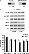
Changes in glutamate receptor levels in the PSD/cytoskeletal fraction. Frontal cortex was dissected from postnatal mice (P11) that had been treated with eNRG1 or vehicle (control) for 9 d. Tissue homogenates were lysed with 1% Triton X-100, fractionated by centrifugation, and separated into the membrane fraction (M2) and PSD/cytoskeletal fraction (P2). A, The fractionation efficiency was examined by Western blotting for PSD95 (a postsynaptic protein) and synaptophysin (a membrane protein). Each lane contained 10 μg of total protein. B, Immunoblots of the PSD/cytoskeletal fraction (P2) were probed with antibodies raised against AMPA receptor subunits (GluA1 and GluA2/3) and NMDA receptor subunits (NR1, NR2A, and NR2B). Representative immunoblots are shown. C, Immunoreactivity levels were measured using densitometry (n = 5 mice each). Results were all normalized to the protein levels in control samples (100%) and plotted. *p < 0.05, t test.
Figure 9.
Figure 9.
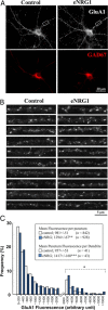
eNRG1 increases surface expression of GluA1 at punctate synaptic structures in cultured GABAergic neurons. Low-density neocortical cultures were prepared from rat embryos (E19) and grown for 2 weeks with or without eNRG1. A, Representative double labeling of surface GluA1 (white) and GAD67 (red) in untreated and eNRG1-treated GABAergic neurons is shown. Dendritic regions (20 μm in length, 20–40 μm away from the soma) were selected randomly from GAD67-positive cells in three control sister cultures (n = 41 cells) and three eNRG1-treated sister cultures (n = 43 cells). B, The top 10 pictures of dendrites showing the higher intensity GluA1 punctuate labeling were selected from each group and arranged from the top based on the signal intensity. C, The frequency of the puncta carrying the indicated range of surface GluA1 intensity was calculated and plotted (n = 842 puncta on 41 dendrites in control cultures; n = 928 puncta on 43 dendrites in eNRG1-treated cultures). **p < 0.01, ***p < 0.001, Student's t test; +p < 0.01, Wilcoxon's test.

References

    1. Abe Y, Namba H, Zheng Y, Nawa H. In situ hybridization reveals developmental regulation of ErbB1–4 mRNA expression in mouse midbrain: implication of ErbB receptors for dopaminergic neurons. Neuroscience. 2009;161:95–110. - PubMed
    1. Barros CS, Calabrese B, Chamero P, Roberts AJ, Korzus E, Lloyd K, Stowers L, Mayford M, Halpain S, Müller U. Impaired maturation of dendritic spines without disorganization of cortical cell layers in mice lacking NRG1/ErbB signaling in the central nervous system. Proc Natl Acad Sci U S A. 2009;106:4507–4512. - PMC - PubMed
    1. Belforte JE, Zsiros V, Sklar ER, Jiang Z, Yu G, Li Y, Quinlan EM, Nakazawa K. Postnatal NMDA receptor ablation in corticolimbic interneurons confers schizophrenia-like phenotypes. Nat Neurosci. 2010;13:76–83. - PMC - PubMed
    1. Buonanno A, Fischbach GD. Neuregulin and ErbB receptor signaling pathways in the nervous system. Curr Opin Neurobiol. 2001;11:287–296. - PubMed
    1. Burden S, Yarden Y. Neuregulins and their receptors: a versatile signaling module in organogenesis and oncogenesis. Neuron. 1997;18:847–855. - PubMed

Publication types

MeSH terms

LinkOut - more resources