

Chondroitinase ABC promotes recovery of adaptive limb movements and enhances axonal growth caudal to a spinal hemisection
- PMID: 21490212
- PMCID: PMC3117673
- DOI: 10.1523/JNEUROSCI.4459-10.2011
Chondroitinase ABC promotes recovery of adaptive limb movements and enhances axonal growth caudal to a spinal hemisection
Abstract
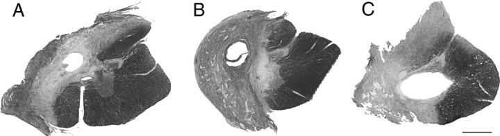
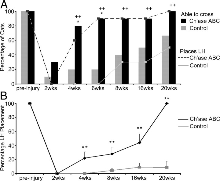
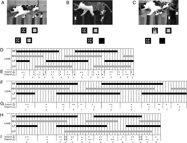
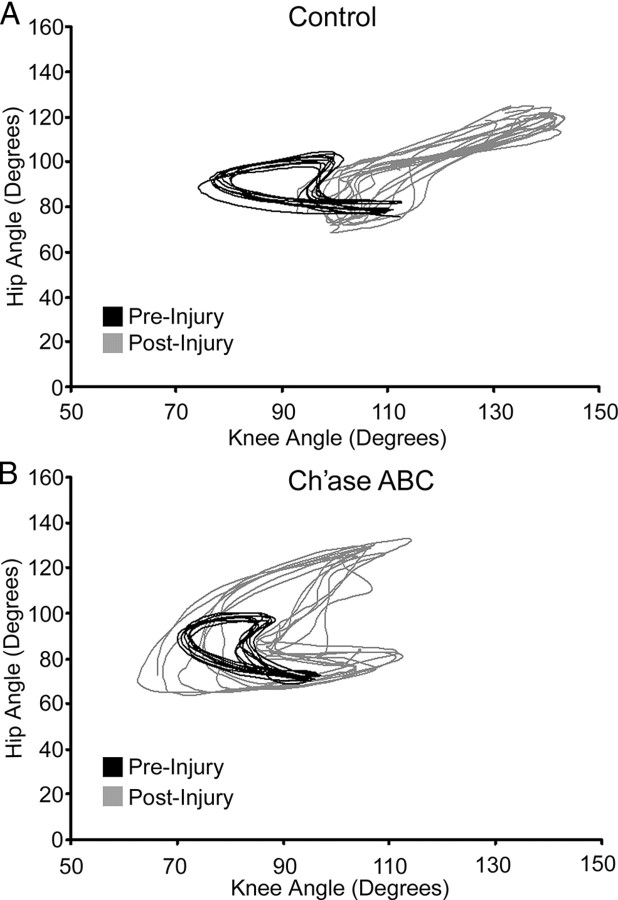
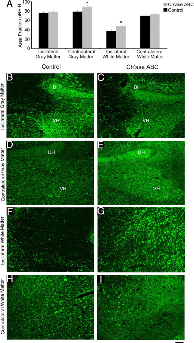
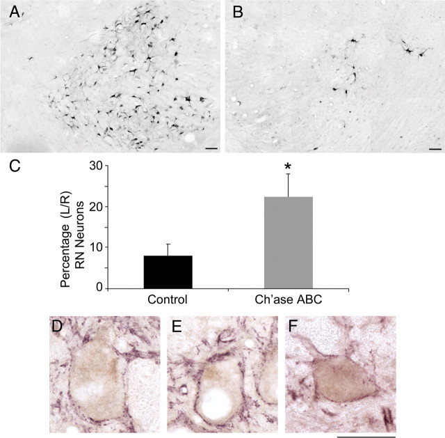
A number of studies have shown that chondroitinase ABC (Ch'ase ABC) digestion of inhibitory chondroitin sulfate glycosaminoglycans significantly enhances axonal growth and recovery in rodents following spinal cord injury (SCI). Further, our group has shown improved recovery following SCI in the larger cat model. The purpose of the current study was to determine whether intraspinal delivery of Ch'ase ABC, following T10 hemisections in adult cats, enhances adaptive movement features during a skilled locomotor task and/or promotes plasticity of spinal and supraspinal circuitry. Here, we show that Ch'ase ABC enhanced crossing of a peg walkway post-SCI and significantly improved ipsilateral hindlimb trajectories and integration into a functional forelimb-hindlimb coordination pattern. Recovery of these complex movements was associated with significant increases in neurofilament immunoreactivity immediately below the SCI in the ipsilateral white (p = 0.033) and contralateral gray matter (p = 0.003). Further, the rubrospinal tract is critical in the normal cat during skilled movements that require accurate paw placements and trajectories like those seen during peg walkway crossing. Rubrospinal connections were assessed following Fluoro-Gold injections, caudal to the hemisection. Significantly more retrogradely labeled right (axotomized) red nucleus (RN) neurons were seen in Ch'ase ABC-treated (23%) compared with control-treated cats (8%; p = 0.032) indicating that a larger number of RN neurons in Ch'ase ABC-treated cats had axons below the lesion level. Thus, following SCI, Ch'ase ABC may facilitate axonal growth at the spinal level, enhance adaptive features of locomotion, and affect plasticity of rubrospinal circuitry known to support adaptive behaviors in the normal cat.
Figures
Similar articles
-
Chondroitinase ABC improves basic and skilled locomotion in spinal cord injured cats.Exp Neurol. 2008 Feb;209(2):483-96. doi: 10.1016/j.expneurol.2007.07.019. Epub 2007 Aug 21. Exp Neurol. 2008. PMID: 17936753 Free PMC article.
-
Axonal regeneration of Clarke's neurons beyond the spinal cord injury scar after treatment with chondroitinase ABC.Exp Neurol. 2003 Jul;182(1):160-8. doi: 10.1016/s0014-4886(02)00052-3. Exp Neurol. 2003. PMID: 12821386
-
Lithium chloride reinforces the regeneration-promoting effect of chondroitinase ABC on rubrospinal neurons after spinal cord injury.J Neurotrauma. 2004 Jul;21(7):932-43. doi: 10.1089/neu.2004.21.932. J Neurotrauma. 2004. PMID: 15307905
-
Transplants and neurotrophic factors increase regeneration and recovery of function after spinal cord injury.Prog Brain Res. 2002;137:257-73. doi: 10.1016/s0079-6123(02)37020-1. Prog Brain Res. 2002. PMID: 12440372 Review.
-
Intervention strategies to enhance anatomical plasticity and recovery of function after spinal cord injury.Adv Neurol. 1997;72:257-75. Adv Neurol. 1997. PMID: 8993704 Review.
Cited by
-
Impact of treatment duration and lesion size on effectiveness of chondroitinase treatment post-SCI.Exp Neurol. 2015 May;267:64-77. doi: 10.1016/j.expneurol.2015.02.028. Epub 2015 Feb 26. Exp Neurol. 2015. PMID: 25725355 Free PMC article.
-
The perineuronal net component of the extracellular matrix in plasticity and epilepsy.Neurochem Int. 2012 Dec;61(7):963-72. doi: 10.1016/j.neuint.2012.08.007. Epub 2012 Aug 29. Neurochem Int. 2012. PMID: 22954428 Free PMC article. Review.
-
Neurological function following intra-neural injection of fluorescent neuronal tracers in rats.Neural Regen Res. 2013 May 15;8(14):1253-61. doi: 10.3969/j.issn.1673-5374.2013.14.001. Neural Regen Res. 2013. PMID: 25206419 Free PMC article.
-
Leukocyte common antigen-related phosphatase is a functional receptor for chondroitin sulfate proteoglycan axon growth inhibitors.J Neurosci. 2011 Oct 5;31(40):14051-66. doi: 10.1523/JNEUROSCI.1737-11.2011. J Neurosci. 2011. PMID: 21976490 Free PMC article.
-
MEK inhibition reduces glial scar formation and promotes the recovery of sensorimotor function in rats following spinal cord injury.Exp Ther Med. 2014 Jan;7(1):66-72. doi: 10.3892/etm.2013.1371. Epub 2013 Oct 29. Exp Ther Med. 2014. PMID: 24348766 Free PMC article.
References
-
- Armstrong DM. Supraspinal contributions to the initiation and control of locomotion in the cat. Prog Neurobiol. 1986;26:273–361. - PubMed
-
- Armstrong DM, Marple-Horvat DE. Role of the cerebellum and motor cortex in the regulation of visually controlled locomotion. Can J Physiol Pharmacol. 1996;74:443–455. - PubMed
-
- Banik NL, Matzelle DC, Gantt-Wilford G, Osborne A, Hogan EL. Increased calpain content and progressive degradation of neurofilament protein in spinal cord injury. Brain Res. 1997;752:301–306. - PubMed
-
- Banik NL, Hogan EL, Powers JM, Whetstine LJ. Degradation of cytoskeletal proteins in experimental spinal cord injury. Neurochem Res. 1982;7:1465–1475. - PubMed
Publication types
MeSH terms
Substances
Grants and funding
LinkOut - more resources
Full Text Sources
Medical
Research Materials
Miscellaneous