. 2011 Apr 13;31(15):5755-63.
doi: 10.1523/JNEUROSCI.5346-10.2011.

Enhanced subcortical spreading depression in familial hemiplegic migraine type 1 mutant mice

Affiliations

Enhanced subcortical spreading depression in familial hemiplegic migraine type 1 mutant mice

Katharina Eikermann-Haerter et al. J Neurosci. .

Abstract

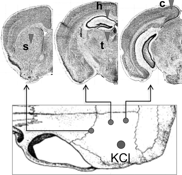
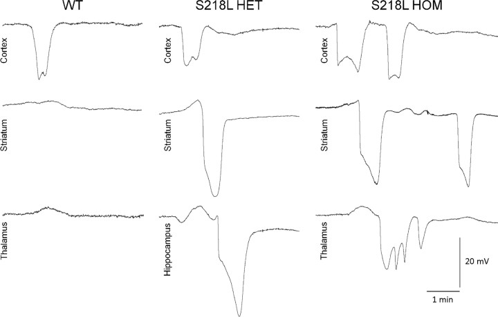
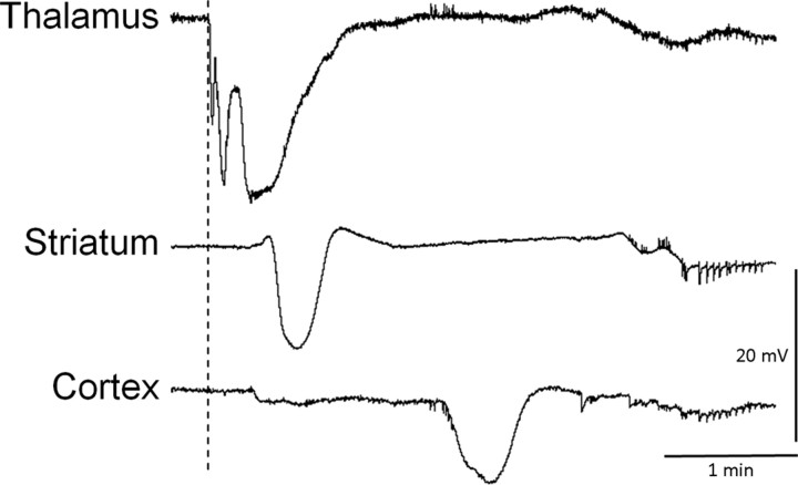
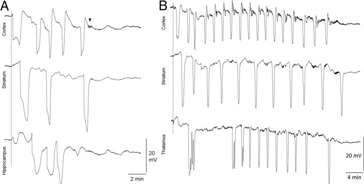
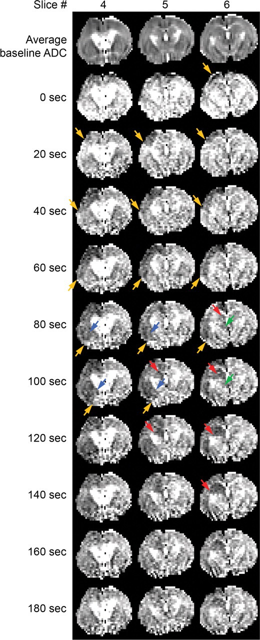
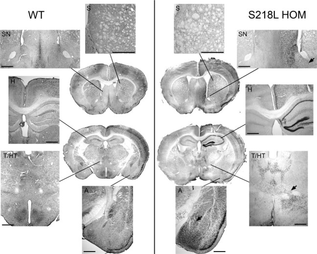
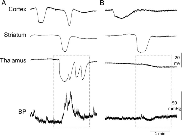
Familial hemiplegic migraine type 1, a monogenic migraine variant with aura, is linked to gain-of-function mutations in the CACNA1A gene encoding Ca(V)2.1 channels. The S218L mutation causes severe channel dysfunction, and paroxysmal migraine attacks can be accompanied by seizures, coma, and hemiplegia; patients expressing the R192Q mutation exhibit hemiplegia only. Familial hemiplegic migraine knock-in mice expressing the S218L or R192Q mutation are highly susceptible to cortical spreading depression, the electrophysiological surrogate for migraine aura, and develop severe and prolonged motor deficits after spreading depression. The S218L mutants also develop coma and seizures and sometimes die. To investigate underlying mechanisms for these symptoms, we used multielectrode electrophysiological recordings, diffusion-weighted magnetic resonance imaging, and c-fos immunohistochemistry to trace spreading depression propagation into subcortical structures. We showed that unlike the wild type, cortical spreading depression readily propagated into subcortical structures in both familial hemiplegic migraine type 1 mutants. Whereas the facilitated subcortical spread appeared limited to the striatum in R192Q, hippocampal and thalamic spread was detected in the S218L mutants with an allele-dosage effect. Both strains exhibited increased susceptibility to subcortical spreading depression and reverberating spreading depression waves. Altogether, these data show that spreading depression propagates between cortex, basal ganglia, diencephalon, and hippocampus in genetically susceptible brains, which could explain the prolonged hemiplegia, coma, and seizure phenotype in this variant of migraine with aura.


Figures

Figure 1.
Figure 1.
Electrophysiological recording sites. SD was triggered by brief topical KCl (300 mm) application onto parietal cortex and recorded with up to four glass micropipettes (arrowheads) placed into cortex (c), striatum (s), hippocampus (h), thalamus (t). [Adapted from Allen Mouse Brain Atlas (Internet), 2009. Seattle: Allen Institute for Brain Science. Available at http://mouse.brain-map.org].
Figure 2.
Figure 2.
Subcortical propagation of cortical SD into hippocampus and thalamus in S218L mutant mice. Representative microelectrode recordings show SD propagation from cortex into striatum and hippocampus or thalamus in HET or HOM S218L mutants but not in WT mice. Thalamic or hippocampal propagation was not found in R192Q HOM mice (see Table 2). Hippocampal SDs were often prolonged compared with other structures. Thalamic SDs were often multiphasic, sometimes coalescing into a prolonged depolarization. The multiphasic waveform may be attributable to multiple corticothalamic propagation pathways arriving sequentially at the thalamic recording site or caused by circling within thalamic nuclei.
Figure 3.
Figure 3.
Reverse propagation of subcortical SD into cortex in S218L HOM mutants. A representative tracing shows a thalamic SD triggered during glass micropipette insertion in S218L HOM mice (dashed line) that preceded slow potential changes in the striatum and cortex, suggesting that SDs can propagate from thalamus to cortex as well.
Figure 4.
Figure 4.
Recurrent cortical and subcortical SDs in S218L HOM mutants. A, B, Representative microelectrode recordings show clusters of SD in cortex, striatum, and hippocampus (A) or thalamus (B) after a single brief epidural KCl application (dashed line) in S218L HOM mice. The arrowhead shows a typical large-amplitude spike burst observed only in the S218L HOM mutant strain. These afterdischarges usually lasted <1 min and started within a few minutes after the recovery of slow potential shift.
Figure 5.
Figure 5.
Diffusion-weighted MRI of SD propagation. Serial magnetic resonance images in a representative S218L HOM mouse show ADC changes during cortical SD (orange) spreading into striatum (blue), hippocampus (red), and dorsomedial thalamus (green). Three consecutive coronal slices are shown over time (acquired at 20 s interval; voxel dimensions: 0.5, 0.2, 0.2 mm). The top row shows average baseline ADC maps to serve as an anatomical reference. Arrows indicate the SD wavefront. Striatal ADC changes corresponding to SD propagation typically occurred in a lateral-to-medial direction, whereas changes in hippocampus propagated in a medial-to-lateral direction. Hippocampal ADC changes persisted longer than in all other regions.
Figure 6.
Figure 6.
Enhanced c-fos expression in cortex and subcortical structures in S218L mutant mice. c-fos immunohistochemistry in coronal sections taken from representative WT and S218L HOM mice 3 h after three consecutive cortical SDs triggered 15 min apart is shown. Coronal sections were taken from two different levels (striatum and hippocampus) in whole brain. Both the number of labeled cells plus staining intensity were increased in S218L mutant brain compared with wild type. Higher-magnification images show that c-fos was expressed throughout cortex in both strains, although this increase appeared more prominent in the S218L HOM mutant. c-fos expression was upregulated in striatum (S) in all mutants (n = 12) but only one WT mouse (n = 6). c-fos upregulation in hippocampus (H) or thalamus (T) was observed in the mutants only. Mutants also showed c-fos upregulation in septal nuclei (SN; 100%), hypothalamus (HT; 50%), and amygdala (A; 100%). c-fos expression was observed in cells bridging contiguous structures such as striatum and amygdala, striatum and septal nuclei, and thalamus and hypothalamus (arrows). Scale bar, 500 μm.
Figure 7.
Figure 7.
Thalamic SDs were associated with transient hypertension. Representative electrophysiological tracings are shown with simultaneous arterial BP recordings in an S218L HOM mutant. A, When an SD propagated into thalamus, transient hypertensive episodes were observed in S218L HOM mice. B, In the absence of thalamic SD, BP increases did not occur, even when SD propagated into the striatum. Hence, hypertensive episodes were never observed in R192Q mutant mice.

Similar articles

Cited by

References

    1. Avoli M, Drapeau C, Louvel J, Pumain R, Olivier A, Villemure JG. Epileptiform activity induced by low extracellular magnesium in the human cortex maintained in vitro. Ann Neurol. 1991;30:589–596. - PubMed
    1. Ayata C, Dunn AK, Gursoy OY, Huang Z, Boas DA, Moskowitz MA. Laser speckle flowmetry for the study of cerebrovascular physiology in normal and ischemic mouse cortex. J Cereb Blood Flow Metab. 2004;24:744–755. - PubMed
    1. Battistini S, Stenirri S, Piatti M, Gelfi C, Righetti PG, Rocchi R, Giannini F, Battistini N, Guazzi GC, Ferrari M, Carrera P. A new CACNA1A gene mutation in acetazolamide-responsive familial hemiplegic migraine and ataxia. Neurology. 1999;53:38–43. - PubMed
    1. Bures J, Fifkova E. The effect of potassium ions liberated from nerve structures invaded by spreading depression on conduction in fibres of the adjacent white matter. Physiol Bohemoslov. 1968;17:405–410. - PubMed
    1. Bures J, Hartman G. Conduction block in capsula interna fibres caused by striatal spreading depression in rats. Experientia. 1967;23:736–737. - PubMed

Publication types

MeSH terms

Substances

Supplementary concepts

LinkOut - more resources