. 2011 Apr 13;31(15):5782-91.
doi: 10.1523/JNEUROSCI.5559-10.2011.

GABA, its receptors, and GABAergic inhibition in mouse taste buds

Affiliations

GABA, its receptors, and GABAergic inhibition in mouse taste buds

Gennady Dvoryanchikov et al. J Neurosci. .

Abstract

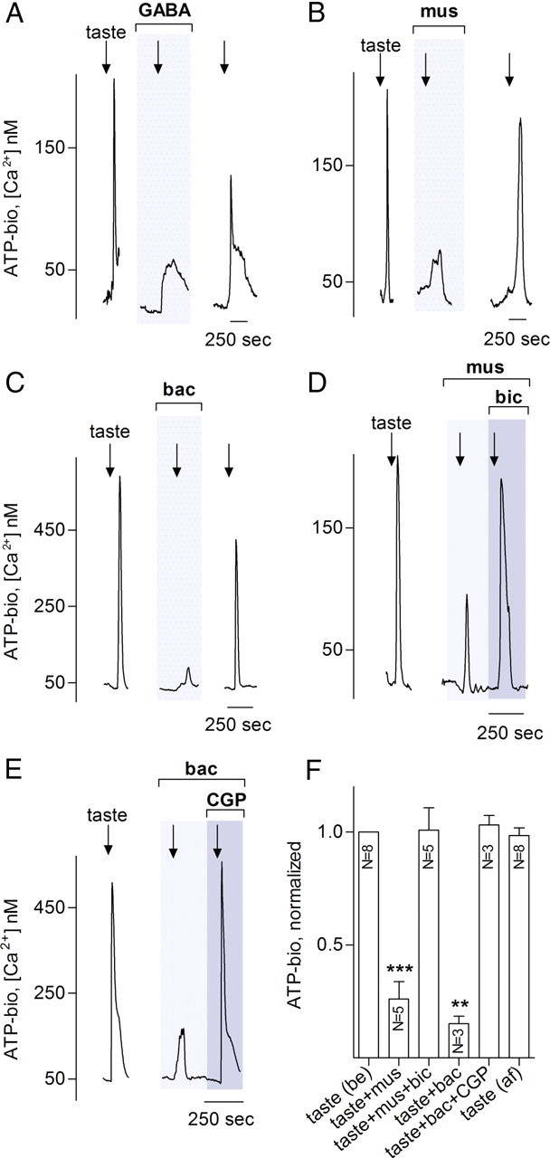
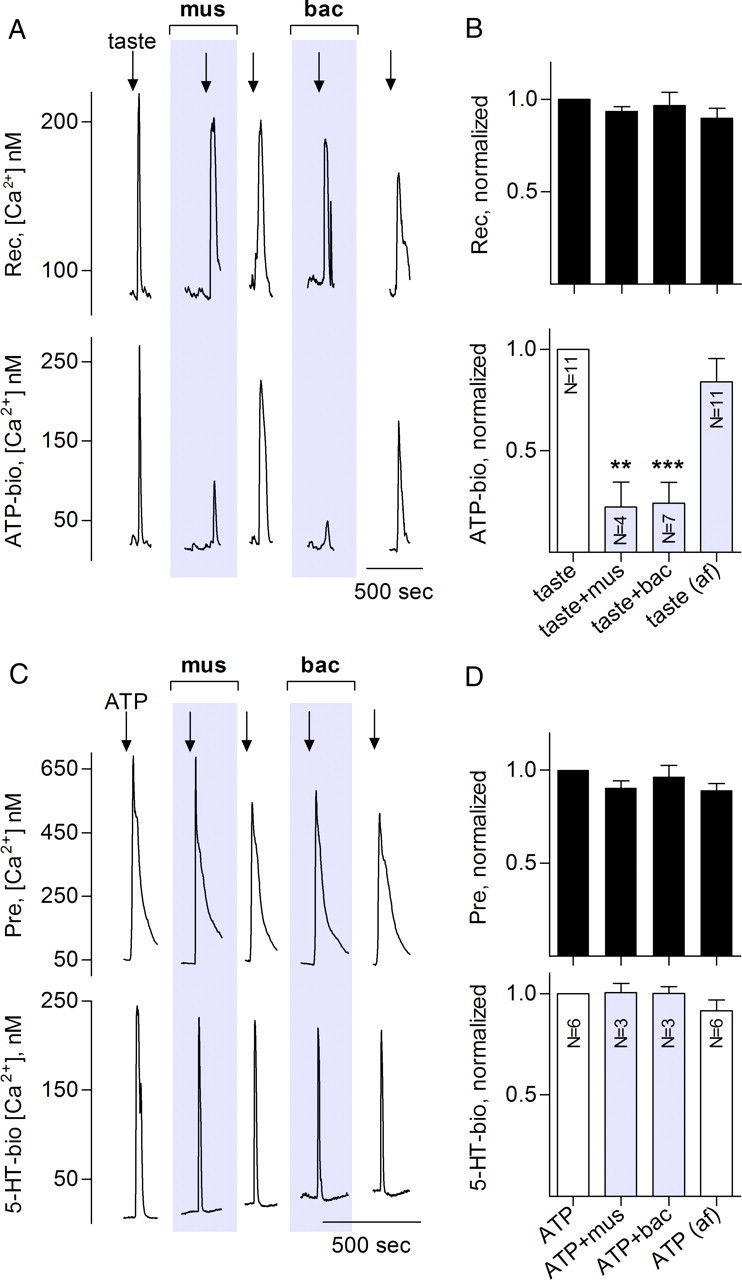
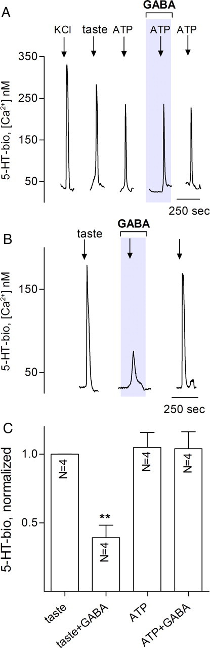
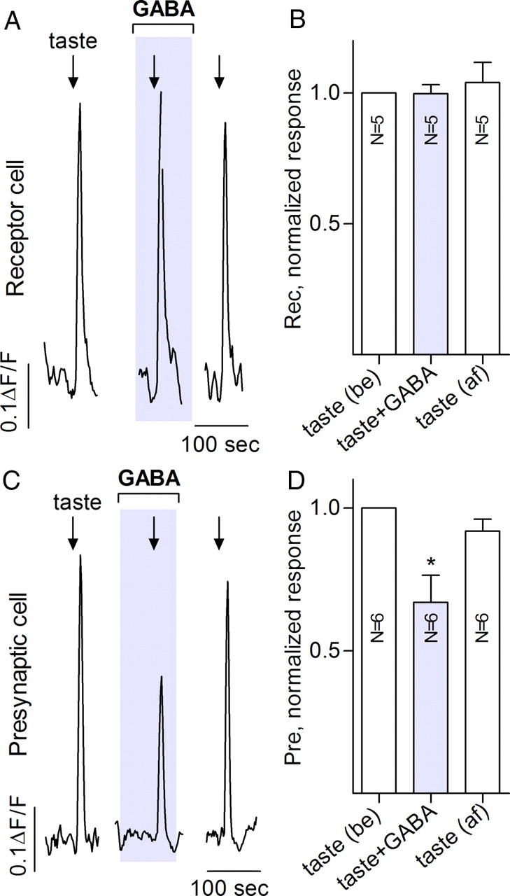
Taste buds consist of at least three principal cell types that have different functions in processing gustatory signals: glial-like (type I) cells, receptor (type II) cells, and presynaptic (type III) cells. Using a combination of Ca2+ imaging, single-cell reverse transcriptase-PCR and immunostaining, we show that GABA is an inhibitory transmitter in mouse taste buds, acting on GABA(A) and GABA(B) receptors to suppress transmitter (ATP) secretion from receptor cells during taste stimulation. Specifically, receptor cells express GABA(A) receptor subunits β2, δ, and π, as well as GABA(B) receptors. In contrast, presynaptic cells express the GABA(A) β3 subunit and only occasionally GABA(B) receptors. In keeping with the distinct expression pattern of GABA receptors in presynaptic cells, we detected no GABAergic suppression of transmitter release from presynaptic cells. We suggest that GABA may serve function(s) in taste buds in addition to synaptic inhibition. Finally, we also defined the source of GABA in taste buds: GABA is synthesized by GAD65 in type I taste cells as well as by GAD67 in presynaptic (type III) taste cells and is stored in both those two cell types. We conclude that GABA is an inhibitory transmitter released during taste stimulation and possibly also during growth and differentiation of taste buds.


Figures

Figure 1.
Figure 1.
GABA inhibits taste-evoked serotonin release from mouse taste buds. Taste buds were isolated from vallate papillae and recorded with biosensors to detect release of serotonin (5-HT). A, Stimulating an isolated taste bud with a bitter–sweet taste mixture (arrows) containing cycloheximide (10 μm), denatonium (1 mm), saccharin (2 mm), and SC45647 (0.1 mm) elicited 5-HT secretion, as shown by the pronounced biosensor response (5-HT-bio). 5-HT secretion was recorded as Δ[Ca2+]i in the serotonin biosensor, measured in nanomolar (see Materials and Methods). Repeating the taste stimulation on the same taste bud in the presence of 10 μm GABA (shaded region, middle trace) showed a marked reduction of 5-HT secretion that was reversed during washout of GABA [GABA did not directly affect 5-HT biosensors (data not shown)]. Similar results were obtained by applying the GABAA receptor agonist muscimol (1 μm) (B) or the GABAB receptor agonist baclofen (C). D, Summary of data from several experiments such as shown in A–C. Taste-evoked serotonin release in the presence of GABA agonists was normalized to serotonin release under control conditions for the same taste bud/biosensor pair (paired Student's t test; *p < 0.05; **p < 0.01). Bars show mean ± SEM. mus, Muscimol (1 μm); bac, baclofen (1 μm); af, after washout of drugs.
Figure 2.
Figure 2.
GABA inhibits taste-evoked ATP release from taste buds. Taste stimulation as in Figure 1 evokes ATP release that is reduced by 10 μm GABA (A), 1 μm muscimol (B), or 1 μm baclofen (C). Traces in A–C show responses from an ATP biosensor that was closely apposed to an isolated taste bud (3 different experiments). Arrows show application of taste mixture, and gray shaded area shows bath perfusion with agonists. D, E, Inhibition of ATP release by muscimol or baclofen is eliminated by specific antagonists to the respective GABA receptors. Thus, 10 μm bicuculline, a GABAA receptor antagonist, rescues taste-evoked ATP secretion even in the presence of muscimol (D) and 10 μm CGP55845, a GABAB receptor antagonist, restores ATP release that was blocked by baclofen. Lightly shaded areas in D and E show bath application of the GABA agonist; and darker shaded area shows perfusion of agonist plus antagonist. Data in D and E verify that the GABA agonists were indeed acting on specific GABA receptors, not generally depressing taste bud cells. F, Summary of data from several experiments as in A–E. Bars show means ± SEM of ATP biosensor responses, normalized as in Figure 1. Paired Student's t test compared each taste-evoked response in the presence of GABAergic drugs to the control evoked response in the same taste bud/biosensor pair (paired Student's t test; ns, no significant difference; **p < 0.01). N, Numbers of taste buds tested; mus, muscimol; bic, bicuculline; bac, baclofen; CGP, CGP55845; be, control before drug application; af, after washout of drugs.
Figure 3.
Figure 3.
Muscimol and baclofen inhibit taste-evoked transmitter release from receptor (type II) but not presynaptic (type III) taste bud cells. Traces in A and C show recordings from an isolated receptor cell (A) or presynaptic cell (B) and its closely apposed biosensor for detecting transmitter release. A, Top trace, Recordings from a receptor cell; bottom trace, ATP biosensor. Repeated taste stimulation (arrows) produces responses in receptor cells and triggers ATP secretion. ATP secretion is significantly and reversibly reduced in presence of 1 μm muscimol or 1 μm baclofen (shaded areas). B, Summary of data from several experiments such as shown in A. Top bars (black) show mean ± SEM for receptor cell responses; bottom bars, concurrent measurements of ATP release (mean ± SEM). Normalization, analyses, and labels as in Figure 1. GABA reduced ATP release (to 34 ± 7%, n = 3; data not shown) similar to that seen for muscimol or baclofen. C, Sequential responses of a paired presynaptic cell and 5-HT biosensor to repeated stimulation with 1 μm ATP (arrows). The presynaptic cell repetitively releases 5-HT, and this release is unaffected by muscimol or baclofen (shaded areas). D, Summary of several experiments on presynaptic cells, similar to summary shown in B for receptor cells. mus, Muscimol; bic, bicuculline; bac, baclofen; be, control before drug application; af, after washout of drugs.
Figure 4.
Figure 4.
In isolated taste buds, GABA interrupts communication between receptor cells and presynaptic cells. Isolated taste buds were sequentially depolarized with KCl, stimulated with tastants and with ATP, in the absence or presence of 10 μm GABA. A, Traces show stimulus-evoked 5-HT release from presynaptic cells within the taste bud (i.e., responses from a 5-HT biosensor apposed to the taste bud). All stimuli (arrows) elicit 5-HT release as shown previously (Huang et al., 2007). GABA has no effect on ATP-evoked 5-HT release. B, Same taste bud, showing effects of GABA on taste-evoked 5-HT release. Here, presynaptic cells are indirectly triggered to release 5-HT, and GABA-mediated inhibition is consistent with GABA acting to reduce ATP release from receptor cells, thereby interfering with cell–cell excitation of presynaptic cells. C, Summary of several experiments as those in A and B. Details of bars and analyses as in Figure 1.
Figure 5.
Figure 5.
Mouse taste buds express distinct GABAA and GABAB receptors in receptor and presynaptic cells. A, RT-PCR on dissected vallate papillae (va), two preparations of cleanly isolated vallate taste buds (tb1, tb2), enzymatically peeled non-taste lingual epithelium (nt), and in parallel, brain (br), and no cDNA (−) control reactions. Expression was tested for several GABAA receptor and one GABAB receptor subunits. RT-PCR for β-actin and the taste-specific marker PLCβ2 on the same samples validated RNA quality and the presence of taste buds only in the first three samples. Data on additional GABAA subunits are presented in supplemental Figure S2 (available at www.jneurosci.org as supplemental material). B, RT-PCR on pools of isolated receptor cells (type II) and presynaptic cells (see Materials and Methods). Three pools of each class were validated by expression of either PLCβ2 or SNAP25 and then were tested for expression of the GABA receptor subunits shown in A. [Although GABAA β1 is expressed in a taste-selective manner (see A), expression was not sufficiently high to permit detection in a few isolated cells.] Control PCRs are as in A. C, Summary of data from expression profiles of receptor and presynaptic cells in B. Incidence of GABA receptor subunits is represented in a modified, overlapping Venn diagram. For example, all three pools of receptor cells but only one of three pools of presynaptic cells expressed GABAB receptors (blue). Receptor cells expressed several different GABAA subunits (warm colors), whereas presynaptic cells express only the β3 isoform (for details, see Results). D, Immunostaining with anti-GABAB1 shows that all receptor cells (visualized as immunopositive for PLCβ2; green) coexpress GABAB1 receptors (red). Scale bar, 20 μm.
Figure 6.
Figure 6.
When tested in a semi-intact preparation, GABA inhibits communication between receptor and presynaptic taste bud cells. Traces show Ca imaging from vallate taste cells in a lingual slice preparation. Receptor cells (A, B) were identified as responding to taste mixture but not 50 mm KCl. Presynaptic cells (C, D) were identified as responding to both the taste mixture and KCl. A, Focally applied taste mixture (arrow) evoked responses in a receptor cell. Taste-evoked receptor cell Ca responses were not significantly affected by bath application of 100 μm GABA (shaded area). B, Summary of data from five experiments as in A. Bars are means ± SEM responses normalized to the group mean for taste responses before (be) GABA. C, In contrast, 100 μm GABA significantly reduces taste-evoked responses recorded from a presynaptic cell. D, Summary of six experiments such as shown in C. Details of bars as in B. Calibration: A, C, 100 s, 0.1 ΔF/F0. be, Control before drug application; af, after washout of drugs.
Figure 7.
Figure 7.
GABA is synthesized and accumulates in both type I and presynaptic (type III) taste bud cells. A, RT-PCR for GAD subtypes in enzymatically peeled epithelium containing taste buds from four oral taste fields: vallate (va), foliate (fo), fungiform (fu), and palate (pa). Both GAD65 and GAD67 are expressed in all fields and little, if at all, in peeled non-taste lingual epithelium (nt). Control reactions include no cDNA (−) and brain cDNA (br). B, Taste bud cells were subjected to single-cell RT-PCR, first to test for expression of NTPDase2, PLCβ2, and SNAP25. Of >50 cells tested, none expressed more than one of these three cell-type markers. The expression of GAD67 and GAD65 in each cell was then tested. The gel shows three representative cells of each type. No expression of GAD65 or GAD67 was detected in receptor cells (Plcβ2+). C, Summary of single-cell profiling of 45 cells as in B shows that GAD65 is only expressed in type I (glial-like) cells, whereas GAD67 is only expressed in presynaptic cells. Receptor (type II) cells do not express either GABA-synthesizing enzyme. D–G, Double immunofluorescence for GABA or its synthetic enzymes and markers for type I, receptor, and presynaptic cells in mouse vallate taste buds. D, GAD65 immunoreactivity (red) is in cells that express NTPDase2 (green), a marker for type I cells. E, GFP fluorescence (green) in taste buds from GAD67–GFP transgenic mice is in cells distinct from cells that are immunoreactive for GAD65 (red). F, GABA immunoreactivity (red) in vallate taste buds from a GAD67–GFP mouse occurs in cells displaying GFP fluorescence (green) as well as in cells that are immunoreactive for NTPDase2 (blue). That is, GABA accumulates only in GAD67-positive type III cells and in NTPDase2-positive type I cells. G, In contrast, GFP fluorescence (green) in taste buds from PLCβ2–GFP transgenic mice does not colocalize with GABA immunostaining, confirming that receptor (PLCβ2+) cells do not synthesize or store GABA.
Figure 8.
Figure 8.
Schematic diagram of GABAergic inhibition in taste buds. The three functional taste cell types are illustrated. Sweet, bitter, or umami stimuli evoke Ca2+ mobilization in receptor cells, leading to ATP secretion. This ATP stimulates both afferent fibers (data not shown) and adjacent presynaptic cells that then secrete 5-HT. GABA, released from presynaptic, type I cells, or both activates GABAA and GABAB receptors (gray and black) on receptor cells and reduces the taste-evoked secretion of ATP. The trigger(s) for GABA secretion and signaling events downstream of GABA receptor activation remain to be elucidated. For clarity, GABA receptors on non-receptor cells are not shown.

Similar articles

Cited by

References

    1. Angulo MC, Le Meur K, Kozlov AS, Charpak S, Audinat E. GABA, a forgotten gliotransmitter. Prog Neurobiol. 2008;86:297–303. - PubMed
    1. Bartel DL, Sullivan SL, Lavoie EG, Sévigny J, Finger TE. Nucleoside triphosphate diphosphohydrolase-2 is the ecto-ATPase of type I cells in taste buds. J Comp Neurol. 2006;497:1–12. - PMC - PubMed
    1. Belelli D, Harrison NL, Maguire J, Macdonald RL, Walker MC, Cope DW. Extrasynaptic GABAA receptors: form, pharmacology, and function. J Neurosci. 2009;29:12757–12763. - PMC - PubMed
    1. Caicedo A, Jafri MS, Roper SD. In situ Ca2+ imaging reveals neurotransmitter receptors for glutamate in taste receptor cells. J Neurosci. 2000;20:7978–7985. - PMC - PubMed
    1. Cao Y, Zhao FL, Kolli T, Hivley R, Herness S. GABA expression in the mammalian taste bud functions as a route of inhibitory cell-to-cell communication. Proc Natl Acad Sci U S A. 2009;106:4006–4011. - PMC - PubMed

Publication types

MeSH terms