Review
. 2011 Jun 8;18(3):R53-77.
doi: 10.1530/ERC-11-0006. Print 2011 Jun.

Tumor-induced osteomalacia

Affiliations
Review

Tumor-induced osteomalacia

William H Chong et al. Endocr Relat Cancer. .

Abstract

Tumor-induced osteomalacia (TIO) is a rare and fascinating paraneoplastic syndrome in which patients present with bone pain, fractures, and muscle weakness. The cause is high blood levels of the recently identified phosphate and vitamin D-regulating hormone, fibroblast growth factor 23 (FGF23). In TIO, FGF23 is secreted by mesenchymal tumors that are usually benign, but are typically very small and difficult to locate. FGF23 acts primarily at the renal tubule and impairs phosphate reabsorption and 1α-hydroxylation of 25-hydroxyvitamin D, leading to hypophosphatemia and low levels of 1,25-dihydroxy vitamin D. A step-wise approach utilizing functional imaging (F-18 fluorodeoxyglucose positron emission tomography and octreotide scintigraphy) followed by anatomical imaging (computed tomography and/or magnetic resonance imaging), and, if needed, selective venous sampling with measurement of FGF23 is usually successful in locating the tumors. For tumors that cannot be located, medical treatment with phosphate supplements and active vitamin D (calcitriol or alphacalcidiol) is usually successful; however, the medical regimen can be cumbersome and associated with complications. This review summarizes the current understanding of the pathophysiology of the disease and provides guidance in evaluating and treating these patients. Novel imaging modalities and medical treatments, which hold promise for the future, are also reviewed.


Conflict of interest statement

Declaration of interest

The authors declare that there is no conflict of interest that could be perceived as prejudicing the impartiality of the research reported.

Figures

Figure 1
Figure 1
Clinical effects of advanced tumor-induced osteomalacia (TIO). The patient in the gown in panel A is depicted standing next to his father. The patient was previously taller than his father, but this is no longer the case. Panel B demonstrates kyphosis and pectus carinatum, which resulted from multiple compression fractures due to osteomalacia. While these findings are the result of advanced osteomalacia, they are strikingly similar to those seen in advanced hyperparathyroidism, as demonstrated by the famous patient reported by Fuller Albright, Captain Martell, shown in panels C and D, who suffered from years of untreated hyperparathyroidism. (Photo of patient and father are reproduced with their permission. Material is reproduced with permission from Albright & Reifenstein (1948)).
Figure 2
Figure 2
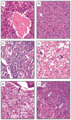
TIO tumor histopathology. (A) This tumor area shows immature mesenchymal cells, with no particular differentiation. There are areas of edema and blood lacunae (arrow). (B) This proliferation is solid, with what seems to be abundant intercellular matrix (arrow). The nuclei are typical with some variation in shape and size. (C) Numerous and irregular vascular structures (arrows), as well as areas of solid proliferation, are seen in this tumor area. (D) This tumor is composed mostly of irregular vascular structures (arrows) embedded in a relatively soft matrix. Variations in size and shapes of the nuclei are evident. (E) Lattice-like areas with ossification (arrow). (F) This photomicrograph was taken from a lung metastasis. The area shows numerous very large osteoclastic-like giant cells (arrows). Note that even though the proliferation is biologically malignant, there are few or no histological signs of malignancy.
Figure 3
Figure 3
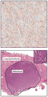
(A) FGF23 immunohistochemistry of lung metastasis of a PMTMCT. (B) The tumor shown in this slide was relatively homogeneous and composed mainly of spindle-shaped cells. Note the relatively benign histological appearance of the neoplasia (inset). FGF23 reactivity was present in almost 100% of the tumor cells.
Figure 4
Figure 4
Summary of the approach to diagnosis and treatment of patients with TIO. Pi, phosphate; %TRP, tubular reabsorption of phosphate; 1,25-D, 1,25-dihydroxyvitamin D.
Figure 5
Figure 5
Subcutaneous TIO tumor detectable on physical examination. Physical examination of a patient with TIO (the same patient shown in Fig. 1) revealed a subcutaneous nodule (A). This nodule was implicated as the culprit tumor by the fact that it was detected on functional imaging, FDG-PET (B). The lesion was visualized on CT scan, which also suggested intralesional calcification (C, arrow). On the low-power view, it can be seen that the tumor was completely contained in the subcutaneous tissue (D), and on a high-power view, it was revealed that the calcification in the lesion was actually ossification and contained areas of lamellar bone (E). TIO resolved after excision.
Figure 6
Figure 6
Multiple imaging modalities may be needed to localize TIO tumors. In this patient, FDG PET/CT revealed multiple areas of increased uptake (A). Octreotide scan only demonstrated a single lesion (B–D). MRI revealed a tumor in the area identified on functional imaging (E). TIO resolved after excision of the lesion.
Figure 7
Figure 7
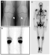
‘Pseudo-reactivation’ of growth plate. X-ray (A) and bone scan (B) of a patient with TIO showing areas of intense tracer uptake and evidence of ‘growth plates’ in a 54-year-old man who had senesced his growth plates decades before. A bone scan of another adult patient with TIO (C) revealed intense tracer uptake at multiple costochondral junctions, creating the nuclear medicine equivalent of an adult rachitic rosary.
Figure 8
Figure 8
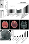
Utility of selective venous sampling in TIO. Selective venous sampling is useful in distinguishing between multiple suspect lesions, as in this patient who had uptake at both the region of the acetabulum (A) and patella (B). Elevated FGF23 in the veins draining the acetabular region (C) identified the lesion in this region as the causative lesion. Selective venous sampling is also useful in identifying lesions in places difficult to image or to approach surgically. The brain shows generalized increased uptake on FDG PET/CT (D) making identification of a lesion in this area difficult. A lesion was seen on octreotide scan (E) and MRI (F). It was felt that this could be a TIO tumor or a meningioma, which are also octreotide positive. Venous sampling (G) demonstrated elevated FGF23 (H), confirming that the lesion was the FGF23-secreting tumor. This material is reproduced from Andreopoulou et al. 2010b with permission of John Wiley & Sons, Inc. r, right; l, left; pop, popliteal; sfv, superficial femoral vein; fem, femoral; prox, proximal; comm, common; int, internal; isch, ischial; mid, middle.

References

    1. ADHR Consortium. Autosomal dominant hypophosphataemic rickets is associated with mutations in FGF23. The ADHR Consortium. Nature Genetics. 2000;26:345–348. doi: 10.1038/81664. - DOI - PubMed
    1. Ahn JM, Kim HJ, Cha CM, Kim J, Yim SG. Oncogenic osteomalacia: induced by tumor, cured by surgery. Oral Surgery, Oral Medicine, Oral Pathology, Oral Radiology, and Endodontics. 2007;103:636–641. doi: 10.1016/j.tripleo.2005.12.027. - DOI - PubMed
    1. Albright F, Reifenstein E. Parathyroid Glands & Metabolic Bone Disease. Vol. 3. Baltimore, MD, USA: The Williams & Wilkins Company; 1948. Clinical hyperparathyroidism; p. 56.
    1. Albright F, Butler AM, Bloomber E. Rickets resistant to vitamin D therapy. American Journal of Diseases of Children. 1937;54:529–547.
    1. Alon U, Hellerstein S. Assessment and interpretation of the tubular threshold for phosphate in infants and children. Pediatric Nephrology. 1994;8:250–251. doi: 10.1007/BF00865491. - DOI - PubMed

Publication types

MeSH terms

Supplementary concepts

LinkOut - more resources