. 2011 Apr 13;3(78):78ra32.
doi: 10.1126/scitranslmed.3002042.

Targeting Nrf2 signaling improves bacterial clearance by alveolar macrophages in patients with COPD and in a mouse model

Affiliations

Targeting Nrf2 signaling improves bacterial clearance by alveolar macrophages in patients with COPD and in a mouse model

Christopher J Harvey et al. Sci Transl Med. .

Abstract

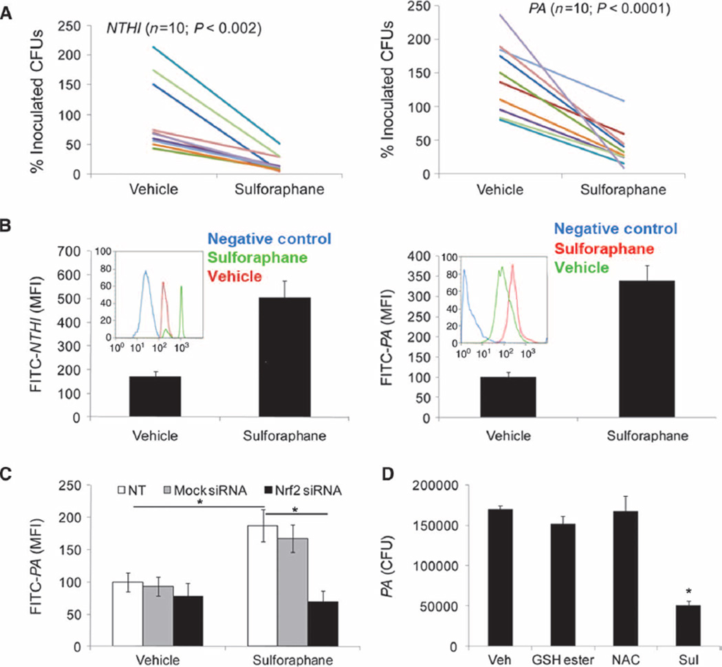
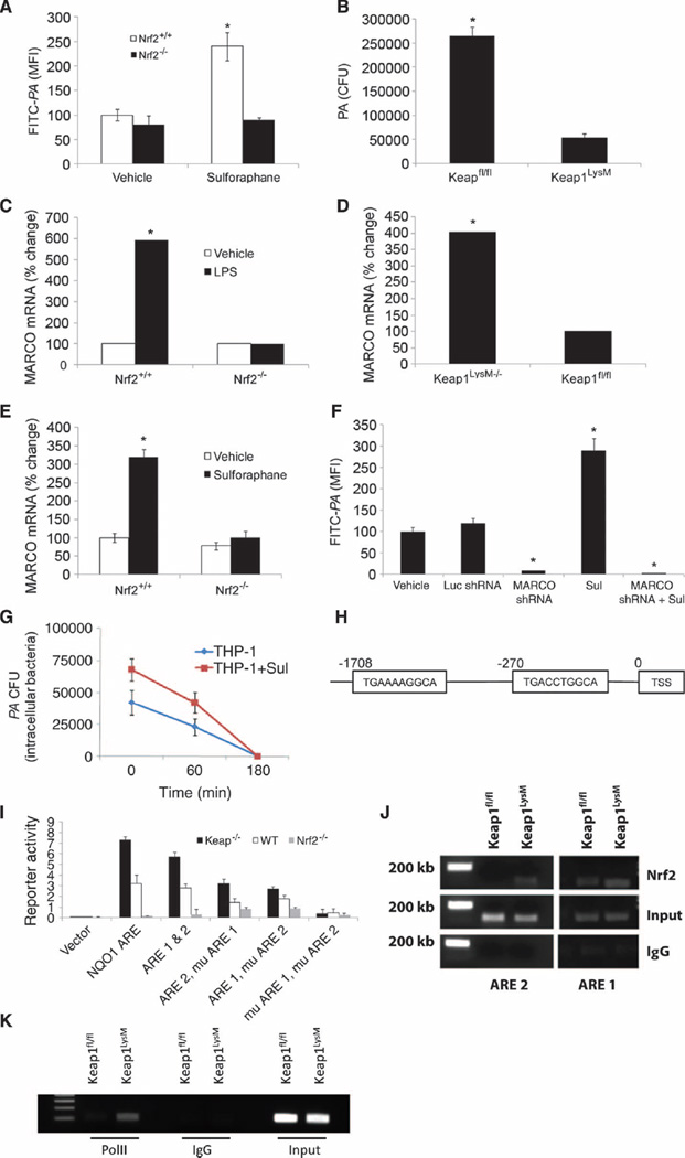
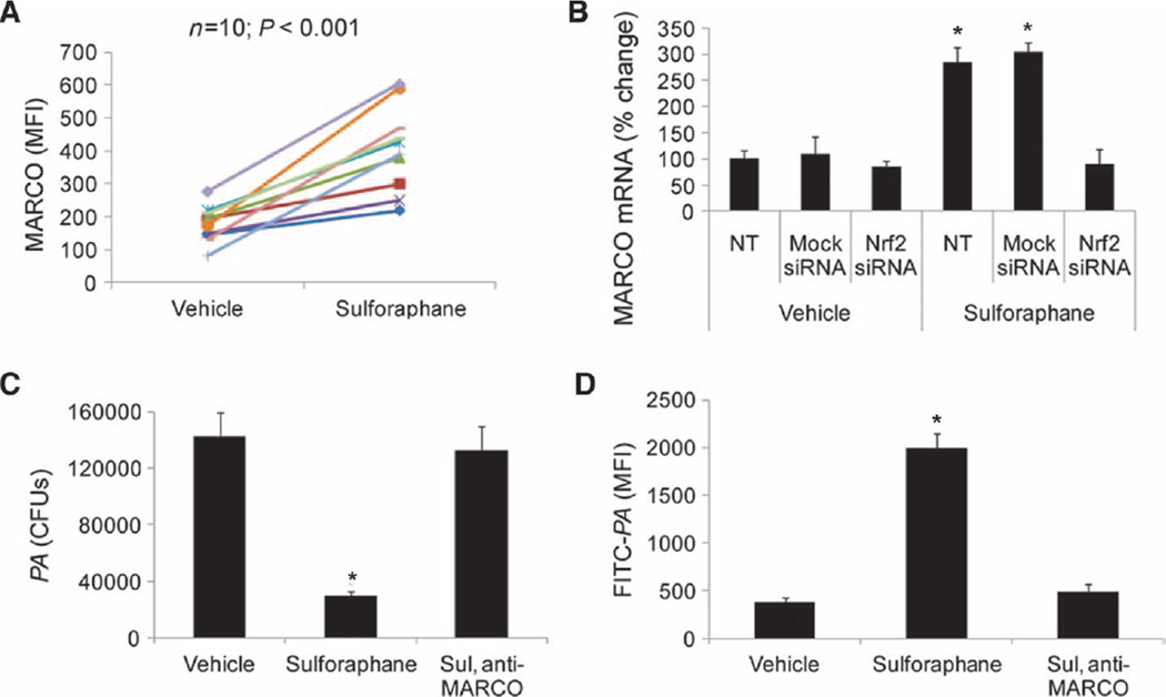
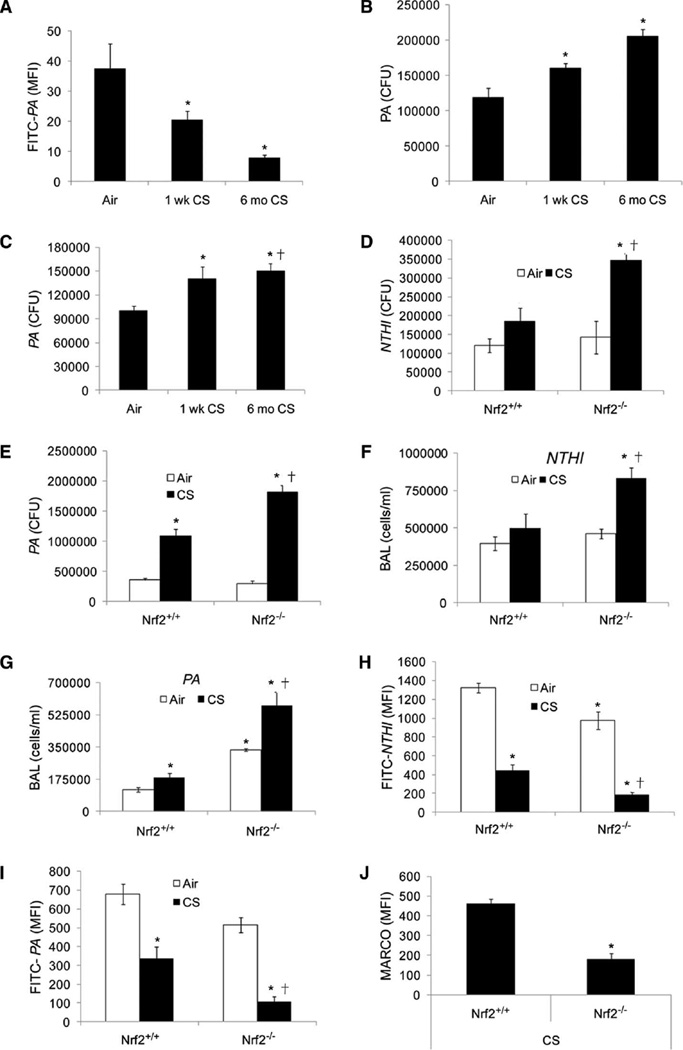
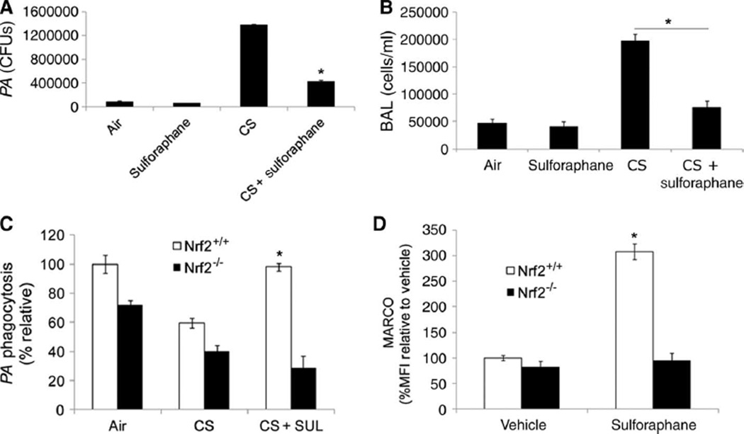
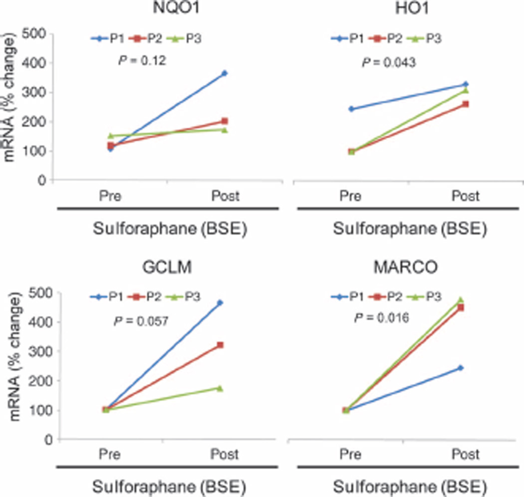
Patients with chronic obstructive pulmonary disease (COPD) have innate immune dysfunction in the lung largely due to defective macrophage phagocytosis. This deficiency results in periodic bacterial infections that cause acute exacerbations of COPD, a major source of morbidity and mortality. Recent studies indicate that a decrease in Nrf2 (nuclear erythroid-related factor 2) signaling in patients with COPD may hamper their ability to defend against oxidative stress, although the role of Nrf2 in COPD exacerbations has not been determined. Here, we test whether activation of Nrf2 by the phytochemical sulforaphane restores phagocytosis of clinical isolates of nontypeable Haemophilus influenza (NTHI) and Pseudomonas aeruginosa (PA) by alveolar macrophages from patients with COPD. Sulforaphane treatment restored bacteria recognition and phagocytosis in alveolar macrophages from COPD patients. Furthermore, sulforaphane treatment enhanced pulmonary bacterial clearance by alveolar macrophages and reduced inflammation in wild-type mice but not in Nrf2-deficient mice exposed to cigarette smoke for 6 months. Gene expression and promoter analysis revealed that Nrf2 increased phagocytic ability of macrophages by direct transcriptional up-regulation of the scavenger receptor MARCO. Disruption of Nrf2 or MARCO abrogated sulforaphane-mediated bacterial phagocytosis by COPD alveolar macrophages. Our findings demonstrate the importance of Nrf2 and its downstream target MARCO in improving antibacterial defenses and provide a rationale for targeting this pathway, via pharmacological agents such as sulforaphane, to prevent exacerbations of COPD caused by bacterial infection.


Conflict of interest statement

Competing interests: S.B., R.K.T., and the Johns Hopkins University hold intellectual property on development of Nrf2-based therapeutics for COPD. Cureveda LLC has licensed this intellectual property. S.B. and R.K.T. have equity in Cureveda, which was co-founded by S.B. and R.K.T. and where they serve on the Scientific Advisory Board. These potential individual and institutional conflicts of interest have been reviewed and managed by the Johns Hopkins University School of Public Health. C.J.H., S.S., X.K., L.Y., R.H.B., D.F.-K., and R.W. declare that they have no competing interests.

Figures

Fig. 1
Fig. 1
Sulforaphane-induced activation of Nrf2 improves phagocytosis and clearance of PA and NTHI by COPD alveolar macrophages. (A) Sulforaphane- or vehicle-treated (16 hours) COPD alveolarmacrophageswere incubated with NTHI or PA. Bacterial load in the culture medium was quantified after 4 hours by serial dilution plating. Data are represented as percent of inoculated CFU remaining in the culture medium for individual patients after vehicle or sulforaphane treatment. (B) COPD alveolar macrophages were incubated with attenuated FITC-labeled NTHI or PA after sulforaphane or vehicle treatment, and phagocytosed bacteria were quantified by flow cytometry after 1 hour. Data are representative histograms from n = 5 patients. Insets: Representative flow cytometry histogram from individual patients. (C) COPD alveolar macrophages transfected with Nrf2 siRNA or ssRNA were treated with sulforaphane or vehicle for 16 hours. Subsequently, macrophages were incubated with FITC-labeled PA and phagocytosed bacteria were quantified by flow cytometry after 1 hour. Data are represented as means ± SEM of MFI (mean fluorescence intensity) (n = 3). (D) GSH ester, NAC, and sulforaphane (Sul)– or vehicle-treated alveolar macrophages isolated from patients with COPD were incubated with PA, and bacterial load in culture medium was quantified after 4 hours. Data are represented as CFU means ± SEM (n = 5 patients).
Fig. 2
Fig. 2
Nrf2 regulates scavenger receptor MARCO expression. (A) BMDMs were derived from Nrf2+/+ and Nrf2−/− mice and treated with vehicle or sulforaphane followed by incubation with FITC-PA. Uptake was quantified by flow cytometry. (B) Alveolar macrophages isolated from Keap1f/f and LysM-Keap1−/− mice were incubated with PA, and bacterial load in culture medium was quantified after 4 hours. Data are represented as CFU means ± SEM. (C) MARCO mRNA expression levels in BMDM isolated from Nrf2+/+ and Nrf2−/− 4 hours after lipopolysaccharide (LPS) or vehicle treatment as measured by microarray analysis. Data are represented as relative fold change compared to vehicle treatment for each genotype. (D) Basal MARCO mRNA expression levels in BMDMs isolated from Keap1f/f and LysM-Keap1−/− as measured by microarray analysis. (E) MARCO mRNA expression levels in BMDMs isolated from Nrf2+/+ and Nrf2−/− 16 hours after sulforaphane or vehicle treatment as measured by qRT-PCR analysis. (F) Phagocytosis of FITC-PA in THP-1 macrophages stably transfected with luciferase (Luc) shRNA or MARCO shRNA and treated with either vehicle or sulforaphane. (G) Intracellular PA (CFUs) in THP-1 macrophage lysates. Lysates were prepared at varying time periods after sulforaphane or vehicle treatment. Data are from an experiment in triplicate. (H) In silico promoter analysis for identification of putative AREs in the 5′ upstream of the transcription start site (TSS) of MARCO gene. (I) Functionality of AREs was evaluated by transfection of luciferase reporter constructs containing ARE1, ARE2; ARE1, mutated (mu) ARE2; ARE2, muARE1; and muARE1, muARE2 into Keap1−/−, Nrf2+/+, and Nrf2−/− MEFs. Data are represented as means ± SEM from three independent experiments. (J) ChIP assay to determine Nrf2 binding to the promoter region of MARCO gene in macrophages derived from LysM-Keap1−/− and Keap1f/f mice. (K) ChIP assay to determine recruitment of RNA PolII (polymerase II) binding to MARCO promoter in macrophages derived from LysM-Keap1−/− and Keap1f/f mice. *P < 0.05.
Fig. 3
Fig. 3
Sulforaphane improves bacterial phagocytic function in COPD alveolar macrophages by Nrf2-dependent up-regulation of MARCO expression. (A) Surface expression of MARCO in COPD alveolar macrophages after sulforaphane or vehicle treatment by flow cytometry. Data are represented as MFI for individual patient with vehicle or sulforaphane treatment. (B) MARCO mRNA expression in COPD alveolar macrophages transfected with mock siRNA, Nrf2 siRNA, or no treatment (NT, untransfected) before sulforaphane treatment. Data are represented as mean percent change compared to vehicle-treated untransfected cells ± SEM (n = 3). *P < 0.05. (C) Bacteria (PA) remaining in the culture medium after blocking MARCO receptor by anti-MARCO antibody in sulforaphane-treated alveolar macrophages. Data are represented as means ± SEM of CFUs in culture medium from each treatment (n=3subjects). (D) Flow analysis of FITC-PA phagocytosis after blocking MARCO receptor by anti-MARCO antibody in sulforaphane-treated alveolar macrophages. Data are represented as means ± SEM of MFI (n = 3 subjects). *P < 0.05, unless otherwise stated.
Fig. 4
Fig. 4
Cigarette smoke exposure impairs bacterial clearance and enhances inflammation in lungs of Nrf2−/− mice when compared to Nrf2+/+ mice. (A) Alveolar macrophages isolated from wild-type mice exposed to filtered air or cigarette smoke (CS) for 1 week or 6 months were incubated with FITC-PA, and uptake was assessed by flow cytometry. Data are represented as means ± SEM of MFI (n = 5 per group). *P < 0.05, compared to air. (B) CFUs in the culture medium of alveolar macrophages isolated from mice exposed to filtered air or cigarette smoke (1 week or 6 months) 4 hours after incubation with PA. Data are represented as means ± SEM of CFUs (n = 5 per group). *P < 0.05, compared to air. (C) Bacterial burden in the lungs of mice exposed to filtered air or cigarette smoke (1 week or 6 months) 4 hours after PA infection. Data are represented as means ± SE of CFUs (n = 5 per group). *P < 0.05, compared to air. (D and E) Bacterial burden in lungs of cigarette smoke (1 week)–or air-exposed Nrf2−/− and Nrf2+/+ mice after 4 hours of NTHI or PA infection. Data are represented as means ± SEM of CFUs (n = 5 per group). *P < 0.05, compared to air-exposed Nrf2+/+; P < 0.05, compared to cigarette smoke–exposed Nrf2+/+. (F and G) Analysis of inflammatory cells in bronchoalveolar lavage (BAL) fluid of cigarette smoke (1 week)– or air-exposed Nrf2+/+ and Nrf2−/− mice 4 hours after NTHI or PA infection. *P < 0.05, compared to air-exposed Nrf2+/+; P < 0.05, compared to cigarette smoke–exposed Nrf2+/+. (H and I) Flow cytometric analysis of FITC-labeled PA or NTHI in alveolar macrophages from Nrf2+/+ and Nrf2−/− mice after cigarette smoke (1 week) or air exposure. *P < 0.05, compared to air-exposed Nrf2+/+; P < 0.05, compared to cigarette smoke–exposed Nrf2+/+. (J) Flow analysis of surface expression of MARCO in alveolar macrophages from Nrf2+/+ and Nrf2−/− mice after cigarette smoke exposure (1 week). Data are represented as means ± SEM of MFI. *P < 0.05, compared to cigarette smoke– exposed Nrf2+/+.
Fig. 5
Fig. 5
Sulforaphane treatment enhances bacterial clearance and reduces inflammation in Nrf2+/+ mice after cigarette smoke exposure. (A) Bacterial burden in lungs of cigarette smoke (1 week) or air-exposed wild-type mice treated with sulforaphane or vehicle 4 hours after PA infection. Data are represented as means ± SEM of CFUs (n = 5 per group). *P < 0.05, compared to cigarette smoke alone. (B) Analysis of inflammatory cells contained in bronchoalveolar lavage (BAL) fluid of cigarette smoke (1 week) or air-exposed, sulforaphane-treated wild-type mice 4 hours after PA infection. Data are represented as means ± SEM (n=5per group). *P < 0.05, compared to cigarette smoke alone. (C) Flow analysis of FITC-labeled PA in alveolar macrophages isolated from sulforaphane (SUL)– or vehicle-treated Nrf2+/+ and Nrf2−/− mice after cigarette smoke (1 week) or air exposure. Data are represented as percent MFI relative to air-exposed Nrf2+/+. (D) Surface expression of MARCO in alveolar macrophages isolated from sulforaphane- or vehicle-treated Nrf2+/+ and Nrf2−/− mice after cigarette smoke (1week) or air exposure as measured by flow cytometry. *P < 0.05, compared to vehicle.
Fig. 6
Fig. 6
Dietary administration of sulforaphane-rich broccoli sprout extract (BSE) enhances MARCO expression in PBMCs. mRNA expression of Nrf2 target genes in PBMCs isolated from three healthy subjects at baseline (Pre) and immediately after 2 weeks (Post) of daily consumption of BSE containing 100 µM sulforaphane. Statistical analysis was conducted by a one-way ANOVA.

References

    1. Barnes PJ. Mediators of chronic obstructive pulmonary disease. Pharmacol. Rev. 2004;56:515–548. - PubMed
    1. Wedzicha JA. Role of viruses in exacerbations of chronic obstructive pulmonary disease. Proc. Am. Thorac. Soc. 2004;1:115–120. - PubMed
    1. Wedzicha JA, Donaldson GC. Exacerbations of chronic obstructive pulmonary disease. Respir. Care. 2003;48:1204–1213. - PubMed
    1. Veeramachaneni SB, Sethi S. Pathogenesis of bacterial exacerbations of COPD. COPD. 2006;3:109–115. - PubMed
    1. Sethi S. Bacteria in exacerbations of chronic obstructive pulmonary disease: Phenomenon or epiphenomenon? Proc. Am. Thorac. Soc. 2004;1:109–114. - PubMed

Publication types

MeSH terms