Review
. 2011 Jul 7;118(1):19-27.
doi: 10.1182/blood-2011-03-325258. Epub 2011 Apr 13.

Fetal hemoglobin in sickle cell anemia

Affiliations
Review

Fetal hemoglobin in sickle cell anemia

Idowu Akinsheye et al. Blood. .

Abstract

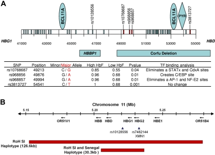
Fetal hemoglobin (HbF) is the major genetic modulator of the hematologic and clinical features of sickle cell disease, an effect mediated by its exclusion from the sickle hemoglobin polymer. Fetal hemoglobin genes are genetically regulated, and the level of HbF and its distribution among sickle erythrocytes is highly variable. Some patients with sickle cell disease have exceptionally high levels of HbF that are associated with the Senegal and Saudi-Indian haplotype of the HBB-like gene cluster; some patients with different haplotypes can have similarly high HbF. In these patients, high HbF is associated with generally milder but not asymptomatic disease. Studying these persons might provide additional insights into HbF gene regulation. HbF appears to benefit some complications of disease more than others. This might be related to the premature destruction of erythrocytes that do not contain HbF, even though the total HbF concentration is high. Recent insights into HbF regulation have spurred new efforts to induce high HbF levels in sickle cell disease beyond those achievable with the current limited repertory of HbF inducers.


Figures

Figure 1
Figure 1
Sickle erythrocytes are a mixture of cells with measurable HbF (F cells) and non-F cells. F cells are long lived, do not acquire the same increment of HbS-induced damage as non-F cells, are less likely to initiate adhesive events, and are associated with protection from sickle vaso-occlusion (left arrow). The heterocellular distribution of HbF in sickle cell anemia, even when total HbF concentrations are high at baseline or in response to hydroxyurea, means that some erythrocytes with no HbF or with suboptimal concentrations of HbF are present. Some of these cells hemolyze intravascularly liberating hemoglobin, which scavenges nitric oxide and contributes to certain vascular complications of this disease (right arrow). This might account for the failure of high HbF that is heterocellularly distributed to protect against all disease complications.
Figure 2
Figure 2
HBB-like gene cluster on chromosome 11 in sickle cell aremia. (A) Results of sequencing 14.1 kb of chromosome 11 in the region of the Corfu deletion. BCL11A binding sites are present between coordinates 44000, 45000, 53000, and 54000. A PYR site is near 54000; rs10128556 is located just 5′ to 47000. Thirty-eight SNPs were found; SNPs marked in red and detailed in the table had a significantly different distribution in 15 patients with HbF more than 11% compared with 15 patients with HbF less than 9%; SNPs in blue had a similar distribution between these groups. Binding sites for BCL11A and PYR are shown in the blue ovals and blue diamond, respectively. Below the coordinates are shown the locations of the β-globin pseudogene (HBBP1) and the Corfu deletion. In the table, minor and major alleles are indicated by black and red, respectively. High HbF and Low HbF represent the major allele frequencies in the 2 groups. Transcription factor binding sites were determined using TFSEARCH and a minimal threshold score of 85.0. Changes in transcription factor binding sites occur in the major allele in the high HbF patients. (B) Runs of SNP homozygosity (RoH) on chromosome 11 in Saudi-Indian and Senegal HBB gene cluster haplotype patients. In the remainder of the genome analyzed with the 610 Illumina array, no other substantial runs of SNP homozygosity were present (A.A., personal communication, March 2011).

References

    1. Rodgers GP, Steinberg MH. Pharmacologic treatment of sickle cell disease and thalassemia: the augmentation of fetal hemoglobin. In: Steinberg MH, Forget BG, Higgs DR, Nagel RL, editors. Disorders of Hemoglobin: Genetics, Pathophysiology, and Clinical Management (1st ed) Cambridge, United Kingdom: Cambridge University Press; 2001. pp. 1028–1051.
    1. Powars D, Weiss JN, Chan LS, Schroeder WA. Is there a threshold level of fetal hemoglobin that ameliorates morbidity in sickle cell anemia? Blood. 1984;63(4):921–926. - PubMed
    1. Platt OS, Brambilla DJ, Rosse WF, et al. Mortality in sickle cell disease. Life expectancy and risk factors for early death. N Engl J Med. 1994;330(23):1639–1644. - PubMed
    1. Steinberg MH, Forget BG, Higgs DR, Weatherall DJ, editors. Disorders of Hemoglobin: Genetics, Pathophysiology, Clinical Management (2nd ed) Cambridge, United Kingdom: Cambridge University Press; 2009.
    1. Kato GJ, Gladwin MT, Steinberg MH. Deconstructing sickle cell disease: reappraisal of the role of hemolysis in the development of clinical subphenotypes. Blood Rev. 2007;21(1):37–47. - PMC - PubMed

Publication types

Substances