. 2011 May;121(5):1871-81.
doi: 10.1172/JCI44393. Epub 2011 Apr 1.

Mutations in 2 distinct genetic pathways result in cerebral cavernous malformations in mice

Affiliations

Mutations in 2 distinct genetic pathways result in cerebral cavernous malformations in mice

Aubrey C Chan et al. J Clin Invest. 2011 May.

Erratum in

  • J Clin Invest. 2012 May 1;122(5):1948

Abstract

Cerebral cavernous malformations (CCMs) are a common type of vascular malformation in the brain that are a major cause of hemorrhagic stroke. This condition has been independently linked to 3 separate genes: Krev1 interaction trapped (KRIT1), Cerebral cavernous malformation 2 (CCM2), and Programmed cell death 10 (PDCD10). Despite the commonality in disease pathology caused by mutations in these 3 genes, we found that the loss of Pdcd10 results in significantly different developmental, cell biological, and signaling phenotypes from those seen in the absence of Ccm2 and Krit1. PDCD10 bound to germinal center kinase III (GCKIII) family members, a subset of serine-threonine kinases, and facilitated lumen formation by endothelial cells both in vivo and in vitro. These findings suggest that CCM may be a common tissue manifestation of distinct mechanistic pathways. Nevertheless, loss of heterozygosity (LOH) for either Pdcd10 or Ccm2 resulted in CCMs in mice. The murine phenotype induced by loss of either protein reproduced all of the key clinical features observed in human patients with CCM, as determined by direct comparison with genotype-specific human surgical specimens. These results suggest that CCM may be more effectively treated by directing therapies based on the underlying genetic mutation rather than treating the condition as a single clinical entity.


Figures

Figure 1
Figure 1. Pdcd10 is required in the endothelium for venous integrity.
(AH) H&E staining of developmental time course of Pdcd10 endothelial knockout. Pdcd10flox/+;Tie2-Cre is shown in AD. Pdcd10flox/–;Tie2-Cre is shown in EH. Close-up images of the cardinal vein at E12.5 are shown in D and H. Asterisks denote the cardinal veins. Circles denote the external jugular veins. (I and J) Echocardiography of hearts from Pdcd10flox/– (I) and Pdcd10flox/–;Tie2-Cre (J) mice at E11.5. Top panels show M-mode images of hearts contracting over time. Bottom panels show waveforms corresponding to blood flow across the atrioventricular valves. s, systole; d, diastole; e, early filling; a, atrial contraction; R, valvular regurgitation. Scale bars: 1 mm (AC and EG); 500 μm (D and H). n ≥ 6 embryos at each age.
Figure 2
Figure 2. PDCD10 differs from CCM2 in downstream signaling.
(A) Phalloidin staining of human microvascular endothelial cells treated with siRNA directed against CCM2, PDCD10, or nonsense control. (B) Quantification of stress fiber response of HMVEC cells. Stress fiber content is determined by adding the total length of stress fibers divided by the total number of cells. Results indicate mean ± SEM and are representative of at least 3 independent experiments. *P < 0.001 versus control or PDCD10; **P = NS versus control. (C) Immunoblot for phospho-myosin light chain-2 (with α-actinin immunoblot as a loading control). Results are representative of 3 independent experiments. Scale bars: 100 μm.
Figure 3
Figure 3. Cavernous malformations result from LOH of either Ccm2 or Pdcd10.
(A) Cavernous malformation (arrow) shown in an H&E-stained section of brain cerebrum from a mouse with induced endothelial knockout of Ccm2. (B) Confirmation of LOH of Ccm2 in 2 mice with loss of Ccm2flox allele by PCR, compared with Pdcd10 wild-type allele as a control. (C) Cavernous malformations (arrows) and a less complex telangiectasia (arrowhead) shown in an H&E-stained section of brain cerebrum from a mouse with induced endothelial knockout of Pdcd10. (D) Confirmation of LOH of Pdcd10 in 2 mice by PCR with loss of Pdcd10flox allele compared with Ccm2 wild-type allele as a control. Samples in B and D were obtained via laser capture microdissection of sectioned mouse brains. Scale bars: 1 mm.
Figure 4
Figure 4. Pathologic analysis of mouse and human PDCD10-associated CCM.
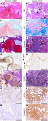
Paired analysis of histologic sections with human tissue on the left and mouse on the right. (A and B) H&E staining revealing back-to-back vascular channels (arrows) and hemosiderin pigment (arrowheads) in surrounding tissues. (C and D) Iron (blue) detected by Prussian blue stain highlights hemosiderin deposits in macrophages and surrounding brain tissue. (E and F) Fibrous matrix deposits (blue) identified by Masson’s trichrome staining with fibrous tissue surrounding vascular channels (arrows) and in surrounding gliotic brain. (G and H) Endothelial staining for CD34 (G) or CD31 (H) is positive in the cells lining the channels. (I and J) Elastin staining shows that vascular channels lack elastic laminae (arrows) unlike normal vessels of similar caliber (arrowhead, inset in I). The fibrous matrix surrounding channels includes laminin (K and L) and collagen IV (M and N). Scale bars: 200 μm.
Figure 5
Figure 5. Pathologic analysis of mouse and human CCM2-associated CCM.
Paired analysis of histologic sections with human tissue on the left and mouse on the right. (A and B) H&E staining revealing back-to-back vascular channels (arrows) and hemosiderin pigment (arrowhead) in surrounding tissues. (C and D) Iron (blue) detected by Prussian blue stain highlights hemosiderin deposits in macrophages and surrounding brain tissue. (E and F) Fibrous matrix deposits (blue) identified by Masson’s trichrome staining with fibrous tissue surrounding vascular channels (arrows) and in surrounding gliotic brain. (G and H) Endothelial staining for CD34 (G) or CD31 (H) is positive in the cells lining the channels. (I and J) Elastin staining shows that vascular channels lack elastic laminae (arrows). The fibrous matrix surrounding channels includes laminin (K and L) and collagen IV (M and N). Scale bars: 200 μm.
Figure 6
Figure 6. Ultrastructural findings in murine cavernous malformations.
(A) Dilated vascular channels are lined by endothelial cells (arrowheads) with associated basal lamina (arrows). (B) Occasional channels have segments with a multilayered appearance (arrows indicate lamellae of endothelium with basal laminae). Tight junctions appear intact (arrowhead). (C) Focal areas of endothelial attenuation are observed (arrow) without apparent gaps or disruption of tight junctions (arrowhead indicates a junctional complex). (D) Channels are separated by loose connective tissue composed mostly of collagen (arrows). (E) Foci of mononuclear inflammatory cells are present (arrows). (F) Hemosiderin-laden macrophages (arrow) are among the inflammatory cells observed. Images are representative of 5 lesions from 3 Pdcd10 mice. Scale bars: 4 μm.
Figure 7
Figure 7. Natural history of murine CCM by MRI — Pdcd10 onsets earlier and is more severe than Ccm2.
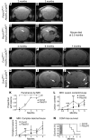
(AD) Live MRI scans of the same Pdcd10flox/+;PDGFb-iCreERT2 mouse at 2 months and 3 months (A and B) and its Pdcd10flox/–;PDGFb-iCreERT2 littermate (C and D). Both mice were given tamoxifen at birth. (EJ) Live MRI scans of the same Ccm2flox/+;PDGFb-iCreERT2 mouse (EG) and its Ccm2flox/–;PDGFb-iCreERT2 littermate (HJ) at 4, 6, and 7 months. Both mice were given tamoxifen at birth. Arrows indicate CCM lesions. (K) Disease penetrance (proportion affected) by age in Ccm2 and Pdcd10 induced knockout mice as assessed by live MRI. (L) Lesion burden assessed as total number of lesions observed on each tomographic view (slice) of the MRI per mouse. (M) Number of complex lesions (lesions with bright cores) per mouse. (N) Kaplan-Meier survival curve of Ccm2 and Pdcd10 induced knockout mice. For KN, n = 11 Ccm2, n = 13 Pdcd10. Data in L and M represent mean ± SEM. *P < 0.01; **P < 0.05; ***P < 0.001. Scale bars: 1 mm.
Figure 8
Figure 8. Convergence of different mechanistic pathways in common pathology.
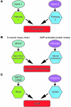
(A) Proposed schema for different genes acting on separate mechanistic pathways, yet ultimately resulting in a common expression of disease. (B) Genetic studies of hypertrophic cardiomyopathy highlight genes that can be grouped broadly into 2 separate mechanistic pathways: sarcomeric proteins such as β-myosin heavy chain (MYH7), and metabolic genes including adenosine monophosphate-activated protein kinase (PRKAG2). (C) Studies of mouse development, cell biology, and signaling suggest that KRIT1 and CCM2 signal through RhoA GTPase, while PDCD10 signals through GCKIII kinases to lead to cavernous malformations.

Similar articles

Cited by

References

    1. Otten P, Pizzolato GP, Rilliet B, Berney J. A propos de 131 cas d’angiomes caverneux (cavernomes) du S.N.C. repérés par l’analyse rétrospective de 24 535 autopsies. Neurochirurgie. 1989;35(2):82–83. - PubMed
    1. Vernooij MW, et al. Incidental findings on brain MRI in the general population. N Engl J Med. 2007;357(18):1821–1828. doi: 10.1056/NEJMoa070972. - DOI - PubMed
    1. Hasegawa T, McInerney J, Kondziolka D, Lee JY, Flickinger JC, Lunsford LD. Long–term results after stereotactic radiosurgery for patients with cavernous malformations. Neurosurgery. 2002;50(6):1190–1197. doi: 10.1097/00006123-200206000-00003. - DOI - PubMed
    1. Chappell PM, Steinberg GK, Marks MP. Clinically documented hemorrhage in cerebral arteriovenous malformations: MR characteristics. Radiology. 1992;183(3):719–724. - PubMed
    1. Burger PC, Scheithauer BW.Tumors of the Central Nervous System . Washington, DC, USA: American Registry of Pathology; 2007.

Publication types

MeSH terms

Substances