Review
. 2011 Jun;79(12):1277-88.
doi: 10.1038/ki.2011.95. Epub 2011 Apr 13.

Intracellular cAMP signaling by soluble adenylyl cyclase

Affiliations
Review

Intracellular cAMP signaling by soluble adenylyl cyclase

Martin Tresguerres et al. Kidney Int. 2011 Jun.

Abstract

Soluble adenylyl cyclase (sAC) is a recently identified source of the ubiquitous second messenger cyclic adenosine 3',5' monophosphate (cAMP). sAC is distinct from the more widely studied source of cAMP, the transmembrane adenylyl cyclases (tmACs); its activity is uniquely regulated by bicarbonate anions, and it is distributed throughout the cytoplasm and in cellular organelles. Due to its unique localization and regulation, sAC has various functions in a variety of physiological systems that are distinct from tmACs. In this review, we detail the known functions of sAC, and we reassess commonly held views of cAMP signaling inside cells.


Conflict of interest statement

DISCLOSURE STATEMENT:

The authors declare they have no conflicts of interest.

Figures
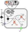
Figure 1
Figure 1
Activation of sAC by HCO3 and Ca2+. Cytosolic sAC can be activated by HCO3 derived from carbonic anhydrase (CA)-dependent hydration of (a) external and (b) metabolic CO2; and/or (c) HCO3 that enters via membrane transporting proteins (purple icon) such as anion exchangers, Na+/HCO3 cotransporters (NBC) or CFTRs. sAC can also be activated by (d) Ca2+ entering the cell via membrane transporters (green icon) such as voltage-dependent Ca2+ channels or potentially via Ca2+ release from the ER or mitochondria (not depicted). (e) HCO3 and Ca2+ can potentially activate sAC in the nucleus. (f) sAC inside mitochondria has been shown to be activated by metabolically generated CO2 via CA. See text for details.
Figure 2
Figure 2
Intracellular cAMP signaling microdomains. cAMP signaling occurs in discrete intracellular compartments such as the membrane vicinity, focal cytoplasm points, mitochondria and the nucleus. Each microdomain contains (1) a source of cAMP (soluble adenylyl cyclase -sAC- or transmembrane adenylyl cyclase -tmAC-); (2) phosphodiesterases (PDE) that act as barriers for cAMP diffusion; and (3) cAMP targets such as protein kinase A (PKA) or Exchange proteins activated by cAMP (EPAC) (not illustrated). tmAC cAMP signaling occurs in response to various extracellular ligands and it requires modulation by G-Protein coupled receptors and heterotrimeric G-protein. The most widely described tmAC-dependent microdomain occurs at the cell membrane, but additional intracellular tmAC-dependent microdomains occur in endosomes after internalization. sAC present throughout the cytoplasm and in organelles such as mitochondria, nucleus, mid-bodies and centrioles define other microdomains. Additional regulation might involve the movement of sAC between compartments. See text for details.
Figure 3
Figure 3
Regulation of V-type H+-ATPase translocation by sAC and AMPK. (1) Extracellular HCO3 enters the cell via transporter proteins (purple icon) or is dehydrated into CO2, a reaction catalyzed by extracellular carbonic anhydrase (CAIV). CO2 would then diffuse into the cell, where it is hydrated into H+ and HCO3 by intracellular carbonic anhydrase (CAII). (2) The elevated intracellular [HCO3] activates sAC (3) to produce cAMP, which promotes (via PKA) (4) the insertion of VHA-containing vesicles into the cell membrane. (5) Membrane inserted VHAs secrete H+, which counteract the original alkalosis. (6) cAMP is hydrolyzed by phosphodiesterase (PDE) into AMP, which can (7) via stimulation of AMPK, inhibit the PKA mediated effects. This hypothetical mechanism, involving sequential activation of PKA and AMPK, could serve as a self-regulating circuit.

References

    1. Braun T, Dods RF. Development of a Mn-2+-sensitive, “soluble” adenylate cyclase in rat testis. Proc Natl Acad Sci U S A. 1975;72:1097–1101. - PMC - PubMed
    1. Braun T. Purification of soluble form of adenylyl cyclase from testes. Methods Enzymol. 1991;195:130–136. - PubMed
    1. Neer EJ. Physical and functional properties of adenylate cyclase from mature rat testis. J Biol Chem. 1978;253:5808–5812. - PubMed
    1. Braun T, Frank H, Dods R, Sepsenwol S. Mn2+-sensitive, soluble adenylate cyclase in rat testis. Differentiation from other testicular nucleotide cyclases. Biochim Biophys Acta. 1977;481:227–235. - PubMed
    1. Forte LR, Bylund DB, Zahler WL. Forskolin does not activate sperm adenylate cyclase. Mol Pharmacol. 1983;24:42–47. - PubMed

LinkOut - more resources