Comparative Study
. 2011 Jul;36(8):1714-28.
doi: 10.1038/npp.2011.52. Epub 2011 Apr 13.

Gender-specific effect of Mthfr genotype and neonatal vigabatrin interaction on synaptic proteins in mouse cortex

Affiliations
Comparative Study

Gender-specific effect of Mthfr genotype and neonatal vigabatrin interaction on synaptic proteins in mouse cortex

Elinor Blumkin et al. Neuropsychopharmacology. 2011 Jul.

Abstract

The enzyme methylenetetrahydrofolate reductase (MTHFR) is a part of the homocysteine and folate metabolic pathways, affecting the methylations of DNA, RNA, and proteins. Mthfr deficiency was reported as a risk factor for neurodevelopmental disorders such as autism spectrum disorder and schizophrenia. Neonatal disruption of the GABAergic system is also associated with behavioral outcomes. The interaction between the epigenetic influence of Mthfr deficiency and neonatal exposure to the GABA potentiating drug vigabatrin (GVG) in mice has been shown to have gender-dependent effects on mice anxiety and to have memory impairment effects in a gender-independent manner. Here we show that Mthfr deficiency interacts with neonatal GABA potentiation to alter social behavior in female, but not male, mice. This impairment was associated with a gender-dependent enhancement of proteins implicated in excitatory synapse plasticity in the female cortex. Reelin and fragile X mental retardation 1 protein (FMRP) levels and membrane GluR1/GluR2 ratios were elevated in wild-type mice treated neonatally with GVG and in Mthfr+/- mice treated with saline, but not in Mthfr+/- mice treated with GVG, compared with control groups (wild type treated with saline). A minor influence on the levels of these proteins was observed in male mice cortices, possibly due to high basal protein levels. Interaction between gender, genotype, and treatment was also observed in the GABA pathway. In female mice, GABA Aα2/gephyrin ratios were suppressed in all test groups; in male mice, a genotype-specific enhancement of GABA Aα2/gephyrin was observed. The lack of an effect on either reln or Fmr1 transcription suggests post-transcriptional regulation of these genes. Taken together, these findings suggest that Mthfr deficiency may interact with neonatal GABA potentiation in a gender-dependent manner to interrupt synaptic function. This may illustrate a possible mechanism for the epigenetic involvement of Mthfr deficiency in neurodevelopmental disorders.


Figures
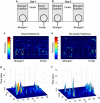
Figure 1
Figure 1
Social behavior analysis. In the social interaction test, mice were introduced on the first day to a three-chambered environment containing a stranger mouse, a central chamber, and a chamber with an empty cage (a). On day 2, mice were tested for preference for social novelty (b), in which mice were introduced to the same three-chambered environment containing the original stranger mouse (Stranger1), a central chamber, and a chamber with an unfamiliar mouse (Stranger2). The duration of time the mice spent at each location of the apparatus is illustrated by a heatmap (maximum value—red; minimal value—blue). An example of social preference, indicated by an increased duration of time spent in Stranger1 chamber compared to empty chamber, is illustrated in (c, upper view) and (d, side view). An example of lack of social preference, indicated by a similar time spent in Stranger1 chamber vs empty chamber, is depicted in (e, upper view) and (f, side view).
Figure 2
Figure 2
Gene–drug effects on preference for social novelty. The distribution patterns of duration in the Stranger1 (S1), center (C), and Stranger2 (S2) chambers of adult female (a) and male (b) mice. n=5–11 (male), n=6–11 (female). χ2-test was used for all analyses. Results are presented as mean±SEM. Female: χ2-tests for each group compared with saline-wild type (Sa-WT), p<0.0001. Male: χ2-test for Sa-WT compared with vigabatrin (GVG)-Mthfr+/− mice, p<0.01.
Figure 3
Figure 3
Effects of gender, genotype, and drug on reelin and Dab1 levels in the cerebral cortex. An example of a reelin immunoblot of the female cortex is shown; the full-length protein, ∼400 kDa, and two products of proteolysis, ∼300 and 180 kDa, were detected by the G-10 anti-reelin antibody. Protein levels were normalized to actin. Reelin levels in female (a) and male (b) cerebral cortex cytoplasmic fractions. Female: reelin 300 kDa, effect of genotype × treatment, F=72.8, p<0.0001; 400 kDa, F=42.4, p<0.0001, male: genotype × treatment, 400 kDa, F=6.9, p<0.02. An example of Dab1 in the male cortex and levels of Dab1 normalized to actin in female and male mice (c). Male: genotype effect, F=26.7, p<0.0001. n=7–8 in each group. Results are presented as mean±SEM.
Figure 4
Figure 4
Effects of gender, genotype, and drug on fragile X mental retardation 1 protein (FMRP) and glutamate receptor (GluR) levels in the cerebral cortex. An example of an FMRP immunoblot of the cytoplasmic fraction from the female cortex (a). Protein levels in cytoplasmic fractions were normalized to actin. FMRP (a) and GluR1 (b) levels in female and male cerebral cortex cytoplasmic fraction. Female FMRP: effect of treatment, F=4.45, p<0.05; effect of treatment × genotype, F=7.7, p<0.02. An example of a GluR1 immunoblot of the cytoplasmic fraction (S2) and the membrane-enriched fraction (LP1), indicating that the GluR1 protein is enriched in the LP1 fraction (c). The GluR1/GluR2 ratio in the LP1 fraction of female and male mice (c). Female GluR1/GluR2 in LP1 fraction; effect of genotype × treatment, F=13.15, p<0.001. n=7–8 in each group. Results are presented as mean±SEM.
Figure 5
Figure 5
Effects of gender and genotype on protein, mRNA, and DNA methylation in the cerebral cortex. Fragile X mental retardation 1 protein (FMRP) levels in the S2 fraction were compared in male and female mice cerebral cortices (a). The glutamate receptor (GluR)1/GluR2 ratio in the plasma membrane-enriched fraction (LP1) fraction in male and female mice was compared (b). n=5 in each group. Real-time PCR analysis of reln and Fmr1 in female cortex (c and d, respectively). n=8–9 in each group. (e) An example of a Southern blot DNA was digested by a combination of the methylation-sensitive enzyme EagI and the methylation-insensitive enzyme EcoRI. DNA was probed with a 569-bp DIG-labeled probe that hybridized to a sequence adjacent to the CGG repeat region in the 5′-UTR of Fmr1. Results are presented as mean±SEM. *p<0.0001; **p<0.05.
Figure 6
Figure 6
Effects of gender, genotype, and drug on γ-aminobutyric acid (GABA) pathway proteins in the cerebral cortex. The levels of GAD65 (a), GABA Aα2 (b), and gephyrin (c) levels normalized to actin levels in the cytoplasmic fractions of female and male cerebral cortices. Female, GAD65: effect of genotype F=6.1, p<0.02. GABA Aα2: effect of vigabatrin (GVG), F=9.01, p<0.01. Gephyrin: effect of GVG, F=16.1, p<0.001, effect of genotype, F=40.16, p<0.0001, genotype × treatment, F=5.03, p<0.05. The GABA Aα2/gephyrin ratio in the plasma membrane-enriched fraction of male and female cerebral cortices (d). Female GABA Aα2/gephyrin: effect of GVG, F=10.01, p<0.005, effect of genotype, F=44.7, p<0.0001, effect of treatment × genotype, F=13.01, p<0.002. Male GABA Aα2/gephyrin: genotype effect, F=14.2, p<0.02, effect of genotype × treatment, F=8.2, p<0.01. n=7–8 in each group. Results are presented as mean±SEM.
Figure 7
Figure 7
Protein methylation in the female brain. (a) An example of S35 radiolabeled protein. A brain homogenate blot of postnatal day P4 Mthfr/−, Mthfr+/+ (wild type (WT)), and P120 WT indicating protein hypomethylation in the Mthfr/− newborns, higher methylated protein status in P4 WT, and the higher level of methylation observed in the P120 WT mice (b). An example of radiolabeled protein of female hippocampus homogenate of all tested groups was shown. Line analysis of protein radiolabeled optical density cortex, hippocampus, and cerebellum (c, d, and e, respectively). Each line represents the average value of three samples.

Similar articles

Cited by

References

    1. Abdulrazzaq YM, Padmanabhan R, Bastaki Salim MA, Ibrahim A, Bener A. Placental transfer of vigabatrin (gamma-vinyl GABA) and its effect on concentration of amino acids in the embryo of TO mice. Teratology. 2001;63:127–133. - PubMed
    1. Abdulrazzaq YM, Padmanabhan R, Bastaki SMA, Ibrahim A, Nurulain M, Shafiullah M. Effect of maternal administration of vigabatrin during late gestation on fetoplacental amino acid profile in the mouse. Reprod Toxicol. 2005;20:549–560. - PubMed
    1. Bagni C, Greenough WT. From mRNP trafficking to spine dysmorphogenesis: the roots of fragile X syndrome. Nat Rev Neurosci. 2005;6:376–387. - PubMed
    1. Beattie EC, Stellwagen D, Morishita W, Bresnahan JC, Ha BK, Von Zastrow M, et al. Control of synaptic strength by glial TNFalpha. Science. 2002;295:2282–2285. - PubMed
    1. Beffert U, Weeber EJ, Durudas A, Qiu S, Masiulis I, Sweatt JD, et al. Modulation of synaptic plasticity and memory by reelin involves differential splicing of the lipoprotein receptor Apoer2. Neuron. 2005;47:567–579. - PubMed

Publication types

MeSH terms