. 2011 May 12;473(7346):221-5.
doi: 10.1038/nature09915. Epub 2011 Apr 13.

Modelling schizophrenia using human induced pluripotent stem cells

Affiliations

Modelling schizophrenia using human induced pluripotent stem cells

Kristen J Brennand et al. Nature. .

Erratum in

  • Nature. 2011 Nov 24;479(7374):556

Abstract

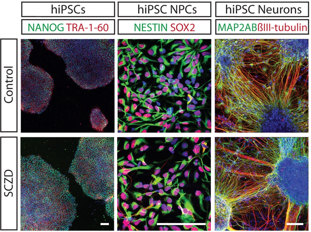
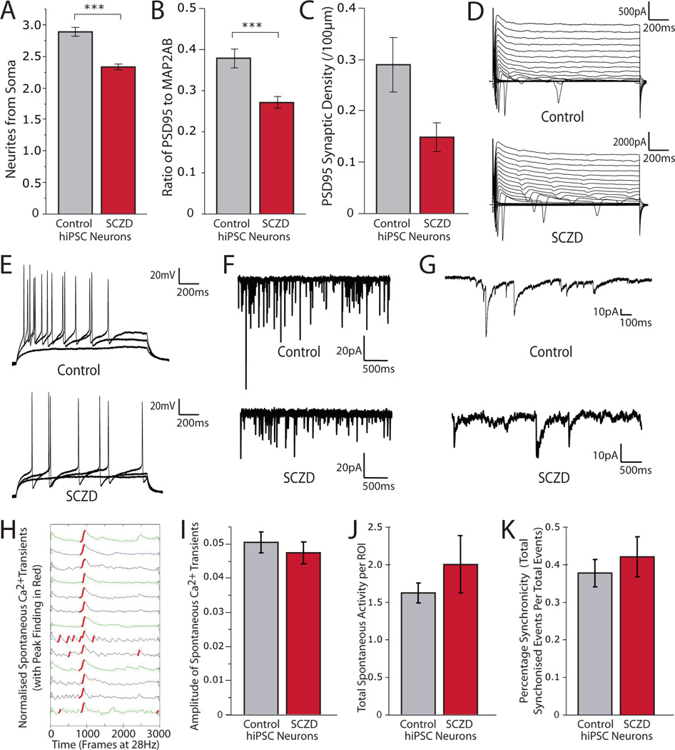
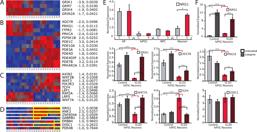
Schizophrenia (SCZD) is a debilitating neurological disorder with a world-wide prevalence of 1%; there is a strong genetic component, with an estimated heritability of 80-85%. Although post-mortem studies have revealed reduced brain volume, cell size, spine density and abnormal neural distribution in the prefrontal cortex and hippocampus of SCZD brain tissue and neuropharmacological studies have implicated dopaminergic, glutamatergic and GABAergic activity in SCZD, the cell types affected in SCZD and the molecular mechanisms underlying the disease state remain unclear. To elucidate the cellular and molecular defects of SCZD, we directly reprogrammed fibroblasts from SCZD patients into human induced pluripotent stem cells (hiPSCs) and subsequently differentiated these disorder-specific hiPSCs into neurons (Supplementary Fig. 1). SCZD hiPSC neurons showed diminished neuronal connectivity in conjunction with decreased neurite number, PSD95-protein levels and glutamate receptor expression. Gene expression profiles of SCZD hiPSC neurons identified altered expression of many components of the cyclic AMP and WNT signalling pathways. Key cellular and molecular elements of the SCZD phenotype were ameliorated following treatment of SCZD hiPSC neurons with the antipsychotic loxapine. To date, hiPSC neuronal pathology has only been demonstrated in diseases characterized by both the loss of function of a single gene product and rapid disease progression in early childhood. We now report hiPSC neuronal phenotypes and gene expression changes associated with SCZD, a complex genetic psychiatric disorder.


Figures

Fig. 1
Fig. 1. Patient-specific hiPSCs, NPCs and neurons
Left. hiPSCs express NANOG (green) and TRA-1-60 (red). DAPI (blue). ×100, scale bar 100 µm. Centre. hiPSC neural progenitor cells (NPCs) express NESTIN (green) and SOX2 (red). DAPI (blue). ×600, scale bar 100 µm. Right. hiPSC neurons express βIII-tubulin (red) and the dendritic marker MAP2AB (green). DAPI (blue). ×200, scale bar 100 µm.
Fig. 2
Fig. 2. Decreased neuronal connectivity in SCZD hiPSC neurons
A. Representative images of control and SCZD hiPSC neurons cotransduced with LV-SYNP-HTG and Rabies-ENVAΔG-RFP, 10 days post rabies transduction. All images were captured using identical laser power and gain settings. βIII-tubulin staining (purple) of the field is shown below each panel. ×400, scale bar 80 µm. B. Graph showing treatment of SCZD hiPSC neurons with Loxapine resulted in a statistically significant improvement in neuronal connectivity. Error bars are s.e., *P < 0.05.
Fig. 3
Fig. 3. Decreased neurites and synaptic protein levels but normal electrophysiological and spontaneous calcium transient activity in SCZD hiPSC neurons
A. Graph showing decreased neurites in SCZD hiPSC neurons. B. Graph showing decreased PSD95 protein relative to MAP2AB for SCZD hiPSC neurons. C. Graph showing a trend of decreased PSD95 synaptic density in SCZD hiPSC neurons. D–G. Electrophysiological characterization. hiPSC neurons cultured on astrocytes show normal sodium and potassium currents when voltage-clamped (D), normal induced action potentials when current-clamped (E), and spontaneous excitatory (F) and inhibitory (G) synaptic activity. H–K. Spontaneous calcium transient imaging. Representative spontaneous Fluo-4AM calcium traces of fluorescent intensity versus time generated from three-month-old hiPSC neurons (H). Graph showing no difference between the spike amplitude of spontaneous calcium transients of control and SCZD hiPSC neurons (I). Graph showing no difference between the total numbers of spontaneous calcium transients per total number of ROIs in cultures of control and SCZD hiPSC neurons (J). Graph showing no change in percentage synchronicity per calcium transient in control and SCZD hiPSC neurons (K). Error bars are SE. Asterisks used as follows: *** p<0.001.
Fig. 4
Fig. 4. RNA expression analysis of control and SCZD hiPSC neurons
A–C. Heat maps showing microarray expression profiles of altered expression of glutamate receptors (A), cAMP signaling (B), and WNT signaling (C) genes in SCZD hiPSC neurons. Fold-change and p-values (diagnosis) provided to the right of each heat map. D. Heat maps showing perturbed expression (highlighted in yellow) of NRG1 and ANK3 in all four SCZD patients, as well as altered expression of ZNF804A, GABRB1, ERBB4, DISC1 and PDE4B in some but not all patients. Fold-change and p-values (diagnosis) provided to the right of each heat map. E. Altered expression of NRG1 is detected in SCZD hiPSC neurons but not in patient fibroblasts, hiPSCs or hiPSC NPCs. F. qPCR validation of altered expression of NRG1, GRIK1, ADCY8, PRKCA, WNT7A, TCF4 and DISC1, as well as response to three weeks of treatment with Loxapine (striped bars) in six-week-old hiPSC neurons. Asterisks used as follows: * p<0.05, ** p<0.01, *** p<0.001.

Comment in

Similar articles

Cited by

References

    1. Sullivan PF, Kendler KS, Neale MC. Schizophrenia as a complex trait: evidence from a meta-analysis of twin studies. Arch Gen Psychiatry. 2003;60:1187–1192. - PubMed
    1. Wong AH, Van Tol HH. Schizophrenia: from phenomenology to neurobiology. Neurosci Biobehav Rev. 2003;27:269–306. - PubMed
    1. Javitt DC, Spencer KM, Thaker GK, Winterer G, Hajos M. Neurophysiological biomarkers for drug development in schizophrenia. Nat Rev Drug Discov. 2008;7:68–83. - PMC - PubMed
    1. Ebert AD, et al. Induced pluripotent stem cells from a spinal muscular atrophy patient. Nature. 2009;457:277–280. - PMC - PubMed
    1. Lee G, et al. Modelling pathogenesis and treatment of familial dysautonomia using patient-specific iPSCs. Nature. 2009;461:402–406. - PMC - PubMed

SI REFERENCES

    1. Association, A. P. Diagnostic and statistical manual of mental disorders: DSM-IV. 3rd ed., rev. edn, Vol. 4th ed. American Psychiatric Press; 1994.
    1. McCarthy SE, et al. Microduplications of 16p11.2 are associated with schizophrenia. Nature genetics. 2009;41:1223–1227. - PMC - PubMed

Publication types

MeSH terms

Associated data