

Hybrid Modality Fusion of Planar Scintigrams and CT Topograms to Localize Sentinel Lymph Nodes in Breast Lymphoscintigraphy: Technical Description and Phantom Studies
- PMID: 21490727
- PMCID: PMC3065894
- DOI: 10.1155/2011/298102
Hybrid Modality Fusion of Planar Scintigrams and CT Topograms to Localize Sentinel Lymph Nodes in Breast Lymphoscintigraphy: Technical Description and Phantom Studies
Abstract
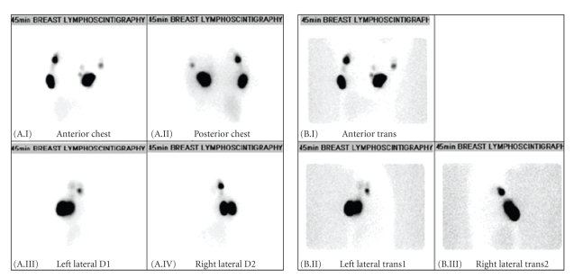
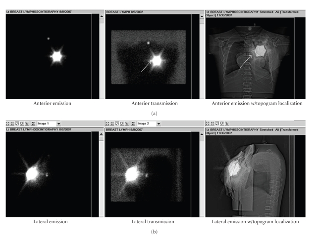
Lymphoscintigraphy is a nuclear medicine procedure that is used to detect sentinel lymph nodes (SLNs). This project sought to investigate fusion of planar scintigrams with CT topograms as a means of improving the anatomic reference for the SLN localization. Heretofore, the most common lymphoscintigraphy localization method has been backlighting with a (57)Co sheet source. Currently, the most precise method of localization through hybrid SPECT/CT increases the patient absorbed dose by a factor of 34 to 585 (depending on the specific CT technique factors) over the conventional (57)Co backlighting. The new approach described herein also uses a SPECT/CT scanner, which provides mechanically aligned planar scintigram and CT topogram data sets, but only increases the dose by a factor of two over that from (57)Co backlighting. Planar nuclear medicine image fusion with CT topograms has been proven feasible and offers a clinically suitable compromise between improved anatomic details and minimally increased radiation dose.
Figures

Similar articles
-
Localisation of sentinel lymph nodes in patients with melanomas by planar lymphoscintigraphic and hybrid SPECT/CT imaging.Nucl Med Rev Cent East Eur. 2012 Aug 27;15(2):101-7. Nucl Med Rev Cent East Eur. 2012. PMID: 22936502
-
Sentinel lymph nodes and planar scintigraphy and SPECT/CT in various types of tumours. Estimation of some factors influencing detection success.Nucl Med Rev Cent East Eur. 2013;16(1):17-25. doi: 10.5603/NMR.2013.0004. Nucl Med Rev Cent East Eur. 2013. PMID: 23677759
-
The clinical value of hybrid sentinel lymphoscintigraphy to predict metastatic sentinel lymph nodes in breast cancer.Nucl Med Mol Imaging. 2015 Mar;49(1):26-32. doi: 10.1007/s13139-014-0298-9. Epub 2014 Oct 17. Nucl Med Mol Imaging. 2015. PMID: 25774235 Free PMC article.
-
The Hybrid SPECT/CT as an additional lymphatic mapping tool in patients with breast cancer.World J Surg. 2008 Sep;32(9):1930-4. doi: 10.1007/s00268-008-9618-5. World J Surg. 2008. PMID: 18478289 Free PMC article. Review.
-
Contribution of SPECT/CT imaging to radioguided sentinel lymph node biopsy in breast cancer, melanoma, and other solid cancers: from "open and see" to "see and open".Q J Nucl Med Mol Imaging. 2014 Jun;58(2):127-39. Q J Nucl Med Mol Imaging. 2014. PMID: 24835289 Review.
Cited by
-
The Usefulness of SPECT/CT in Sentinel Node Mapping of Early Stage Breast Cancer Patients Showing Negative or Equivocal Findings on Planar Scintigraphy.Asia Ocean J Nucl Med Biol. 2018 Spring;6(2):80-89. doi: 10.22038/aojnmb.2018.10720. Asia Ocean J Nucl Med Biol. 2018. PMID: 29998140 Free PMC article.
-
Detection of Sentinel Lymph Nodes in Gynecologic Tumours by Planar Scintigraphy and SPECT/CT.Mol Imaging Radionucl Ther. 2012 Aug;21(2):47-55. doi: 10.4274/Mirt.236. Epub 2012 Aug 1. Mol Imaging Radionucl Ther. 2012. PMID: 23486989 Free PMC article.
-
Evaluation of sentinel lymph node localization in malignant melanoma by preoperative semiconductor gamma camera and planar lymphoscintigraphy.J Appl Clin Med Phys. 2023 Aug;24(8):e14077. doi: 10.1002/acm2.14077. Epub 2023 Jun 26. J Appl Clin Med Phys. 2023. PMID: 37357570 Free PMC article.
References
-
- Krynyckyi BR, Kim CK, Goyenechea MR, Chan PT, Zhang Z-Y, Machac J. Clinical breast lymphoscintigraphy: optimal techniques for performing studies, image atlas, and analysis of images. Radiographics. 2004;24(1):121–139. - PubMed
-
- Krynyckyi BR, Miner M, Ragonese JM, Firestone M, Kim CK, Machac J. Technical aspects of performing lymphoscintigraphy: optimization of methods used to obtain images. Clinical Nuclear Medicine. 2000;25(12):978–985. - PubMed
-
- Mar MV, Dickinson RL, Erwin WD, Wendt RE., III Optimal 57Co flood source activity and acquisition time for lymphoscintigraphy localization images. Journal of Nuclear Medicine Technology. 2008;36(2):82–87. - PubMed
-
- Krynyckyi BR, Chun H, Hyun HK, Eskandar Y, Kim CK, Machac J. Factors affecting visualization rates of internal mammary sentinel nodes during lymphoscintigraphy. Journal of Nuclear Medicine. 2003;44(9):1387–1393. - PubMed
LinkOut - more resources
Full Text Sources