

Reduction of collimator correction artefacts with bayesian reconstruction in spect
- PMID: 21490730
- PMCID: PMC3065867
- DOI: 10.1155/2011/630813
Reduction of collimator correction artefacts with bayesian reconstruction in spect
Abstract
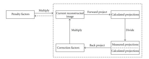
Poor resolution of single photon emission computed tomography (SPECT) has degraded its use in clinical practice. Collimator correction has been shown to improve the reconstructed resolution, but the correction can generate ringing artefacts, which lower image quality. This paper investigates whether Bayesian reconstruction methods could reduce these artefacts. We have applied and tested three Bayesian reconstruction methods: smoothing prior, median root prior, and anatomical prior. To demonstrate the efficacy of these methods, we compared their physical and visual performance both in phantom and patient studies. All the three Bayesian reconstruction methods reduced the collimator correction artefacts. Images reconstructed using the smoothing prior and the median root prior had slightly lower contrast than the standard reconstruction with collimator correction, whereas the anatomical prior produced images with good resolution and contrast.
Figures


Similar articles
-
Correction of photon attenuation and collimator response for a body-contouring SPECT/CT imaging system.J Nucl Med. 2005 May;46(5):868-77. J Nucl Med. 2005. PMID: 15872362 Clinical Trial.
-
Improved quantification in multiple-pinhole SPECT by anatomy-based reconstruction using microCT information.Eur J Nucl Med Mol Imaging. 2011 Jan;38(1):153-65. doi: 10.1007/s00259-010-1627-6. Epub 2010 Sep 30. Eur J Nucl Med Mol Imaging. 2011. PMID: 20882279
-
Transmission imaging for nonuniform attenuation correction using a three-headed SPECT camera.J Nucl Med. 1998 Jun;39(6):1105-10. J Nucl Med. 1998. PMID: 9627354
-
Pinhole single-photon emission tomography reconstruction based on median root prior.Eur J Nucl Med Mol Imaging. 2003 Feb;30(2):217-21. doi: 10.1007/s00259-002-1015-y. Epub 2002 Nov 8. Eur J Nucl Med Mol Imaging. 2003. PMID: 12552339
-
Comparison of multi-ray and point-spread function based resolution recovery methods in pinhole SPECT reconstruction.Nucl Med Commun. 2006 Oct;27(10):823-7. doi: 10.1097/01.mnm.0000237993.83066.0b. Nucl Med Commun. 2006. PMID: 16969266
Cited by
-
Monte carlo study of the effect of collimator thickness on T-99m source response in single photon emission computed tomography.World J Nucl Med. 2012 May;11(2):70-4. doi: 10.4103/1450-1147.103419. World J Nucl Med. 2012. PMID: 23372440 Free PMC article.
-
Validation of Monte Carlo 131 I radiopharmaceutical dosimetry workflow using a 3D-printed anthropomorphic head and neck phantom.Med Phys. 2022 Aug;49(8):5491-5503. doi: 10.1002/mp.15699. Epub 2022 Jun 6. Med Phys. 2022. PMID: 35607296 Free PMC article.
-
Comparison of standardized uptake values between 99mTc-HDP SPECT/CT and 18F-NaF PET/CT in bone metastases of breast and prostate cancer.EJNMMI Res. 2019 Jan 24;9(1):6. doi: 10.1186/s13550-019-0475-z. EJNMMI Res. 2019. PMID: 30680469 Free PMC article.
-
Accuracy of patient-specific I-131 dosimetry using hybrid whole-body planar-SPECT/CT I-123 and I-131 imaging.EJNMMI Phys. 2024 Jun 20;11(1):50. doi: 10.1186/s40658-024-00657-9. EJNMMI Phys. 2024. PMID: 38898326 Free PMC article.
-
Verification of reprojected planar images generated from a ring-configured cadmium zinc telluride gamma camera in scintigraphy for diagnosing transthyretin cardiac amyloidosis.Eur Heart J Imaging Methods Pract. 2024 May 31;2(1):qyae051. doi: 10.1093/ehjimp/qyae051. eCollection 2024 Jan. Eur Heart J Imaging Methods Pract. 2024. PMID: 39224107 Free PMC article.
References
-
- Lau YH, Hutton BF, Beekman FJ. Choice of collimator for cardiac SPET when resolution compensation is included in iterative reconstruction. European Journal of Nuclear Medicine. 2001;28(1):39–47. - PubMed
-
- Gifford HC, King MA, Glenn Wells R, Hawkins WG, Narayanan MV, Pretorius PH. LROC analysis of detector-response compensation in SPECT. IEEE Transactions on Medical Imaging. 2000;19(5):463–473. - PubMed
-
- Frey EC, Gilland KL, Tsui BMW. Application of task-based measures of image quality to optimization and evaluation of three-dimensional reconstruction-based compensation methods in myocardial perfusion SPECT. IEEE Transactions on Medical Imaging. 2002;21(9):1040–1050. - PubMed
-
- He B, Du Y, Song X, Segars WP, Frey EC. A Monte Carlo and physical phantom evaluation of quantitative In-111 SPECT. Physics in Medicine and Biology. 2005;50(17):4169–4185. - PubMed
-
- DePuey EG, Gadiraju R, Clark J, Thompson L, Anstett F, Shwartz SC. Ordered subset expectation maximization and wide beam reconstruction "half-time" gated myocardial perfusion SPECT functional imaging: a comparison to "full-time" filtered backprojection. Journal of Nuclear Cardiology. 2008;15(4):547–563. - PubMed
LinkOut - more resources
Full Text Sources
